
Page 391 - Concise Pathology for Exam Preparation ( PDFDrive )
P. 391
376 SECTION II Diseases of Organ Systems
Clinical course of pneumoconiosis
• Simple coal worker’s pneumoconiosis usually has a benign course; PMF may lead to
cor pulmonale in a few patients.
• Coal dust exposure increases the incidence of chronic bronchitis and emphysema.
Predisposing factors implicated in the development of PMF:
• Older age of the miners
• Amount and duration of exposure to coal dust
• Coexisting tuberculosis
• Coexisting silicosis may further damage the lungs by the following mechanisms:
• Free radical generation (reactive oxygen species that damage the lung parenchyma)
• Release of chaemotactic factors, which induce infiltration of inflammatory cells
into pulmonary tissue
• Release of fibrogenic cytokines—IL-1, TNF and PDGF—which cause healing by
fibrosis
3. Silicosis (knife grinder’s lung disease)
(a) Most prevalent chronic occupational lung disease
(b) Silica has two forms: crystalline and amorphous; the crystalline forms (quartz, cristo-
balite and tridymite) are more fibrogenic and toxic than the noncrystalline forms
(c) Prolonged exposure leads to nodular fibrosing pneumoconiosis
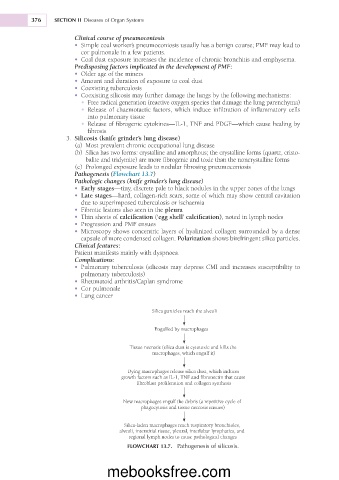
Pathogenesis (Flowchart 13.7)
Pathologic changes (knife grinder’s lung disease)
• Early stages—tiny, discrete pale to black nodules in the upper zones of the lungs
• Late stages—hard, collagen-rich scars, some of which may show central cavitation
due to superimposed tuberculosis or ischaemia
• Fibrotic lesions also seen in the pleura
• Thin sheets of calcification (‘egg shell’ calcification), noted in lymph nodes
• Progression and PMF ensues
• Microscopy shows concentric layers of hyalinized collagen surrounded by a dense
capsule of more condensed collagen. Polarization shows birefringent silica particles.
Clinical features:
Patient manifests mainly with dyspnoea.
Complications:
• Pulmonary tuberculosis (silicosis may depress CMI and increases susceptibility to
pulmonary tuberculosis)
• Rheumatoid arthritis/Caplan syndrome
• Cor pulmonale
• Lung cancer
Silica particles reach the alveoli
Engulfed by macrophages
Tissue necrosis (silica dust is cytotoxic and kills the
macrophages, which engulf it)
Dying macrophages release silica dust, which induces
growth factors such as IL-1, TNF and fibronectin that cause
fibroblast proliferation and collagen synthesis
New macrophages engulf the debris (a repetitive cycle of
phagocytosis and tissue necrosis ensues)
Silica-laden macrophages reach respiratory bronchioles,
alveoli, interstitial tissue, pleural, interlobar lymphatics, and
regional lymph nodes to cause pathological changes
FLOWCHART 13.7. Pathogenesis of silicosis.
mebooksfree.com