
Page 395 - Concise Pathology for Exam Preparation ( PDFDrive )
P. 395
380 SECTION II Diseases of Organ Systems
(c) Asbestosis (particularly, when coupled with smoking)
(d) Nickel, chromates, coal, mustard and arsenic
3. Indoor air pollutants (eg, radon) have been implicated.
4. Role of molecular genetics:
(a) Loss of tumour suppressor genes like P53, RB1 and inactivation of CDK in-
hibitor P 16 are seen equally in adeno and squamous cell carcinoma. P53 and RB1
mutations are common in small cell carcinoma as well. Small cell carcinomas also
commonly demonstrate amplification of genes of MYC family.
(b) Gain of function mutations involving the growth factor receptor signalling path-
ways (genes encoding receptor tyrosine kinases, eg, EGFR, ALK, ROS, MET
and RET), amplifications in epidermal growth factor receptor (EGFR) gene and
mutations in KRAS are typically seen in patients with adenocarcinoma.
(c) Allelic losses on short arm of chromosomes 3, 9 and 17 may precede invasion in
squamous cell carcinoma.
5. Scarring
(a) Scars are most often encountered in the vicinity of adenocarcinomas.
(b) In most cases, scar is a desmoplastic response to tumour; occasionally, scar may
precede carcinoma (old infarcts, foreign bodies, wounds and granulomatous
inflammation).
Four types of Precursor Lesions are recognized:
1) Squamous dysplasia and carcinoma in situ
2) Atypical adenomatous hyperplasia (small , 5 mm lesions, solitary or multiple, composed
of dysplastic pneumocytes lining fibrosed alveolar walls)
3) Adenocarcinoma in situ (formerly called bronchioloalveolar carcinoma it is a lesion
smaller than 3 cm. It is constituted by dysplastic cells which grow along alveolar septa.)
4) Diffuse idiopathic pulmonary neuroendocrine cell hyperplasia.
Morphology
• Most common location is the hilar region.
• Most lesions arise from 1st, 2nd and 3rd order bronchi; few arise from peripheral al-
veolar septal cells and terminal bronchioles.
• Peripheral tumours are usually adenocarcinomas.
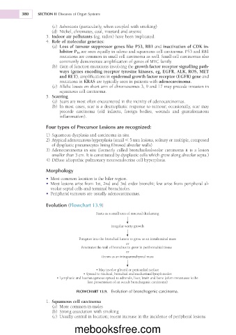
Evolution (Flowchart 13.9)
Starts as a small area of mucosal thickening
Irregular warty growth
Fungates into the bronchial lumen to grow as an intraluminal mass
or
Penetrates the wall of bronchus to grow in peribronchial tissue
or
Grows as an intraparenchymal mass
• May involve pleural or pericardial surface
• Spread to tracheal, bronchial and mediastinal lymph nodes
• Lymphatic and haematogenous spread to adrenals, liver, brain and bone (often metastases is the
first presentation of an occult bronchogenic carcinoma)
FLOWCHART 13.9. Evolution of bronchogenic carcinoma.
1. Squamous cell carcinoma
(a) More common in males
(b) Strong association with smoking
(c) Usually central in location; recent increase in the incidence of peripheral lesions
mebooksfree.com