
Page 396 - Concise Pathology for Exam Preparation ( PDFDrive )
P. 396
13 The Lung 381
(d) Well-differentiated lesions show minimal atypia, intercellular bridges as well as abun-
dant keratin (keratinization is seen as numerous keratin pearls as well as individual
cell keratinization. Squamous cells with intracellular keratin demonstrate abundant
dense eosinophilic cytoplasm).
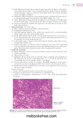
(e) Moderately differentiated lesions show moderate atypia, individual cell keratinization,
occasional keratin pearl, if any and fewer intercellular bridges (Fig. 13.6).
(f) Poorly differentiated lesions are focally keratinized (do not demonstrate keratin easily)
and show severe atypia. These lesions are difficult to recognize as squamous in origin.
(g) Squamous metaplasia, dysplasia and squamous cell carcinoma in situ may be seen
in the adjacent tissue.
2. Adenocarcinoma. It is of two types:
(a) Most common carcinoma in females and nonsmokers
(b) Peripheral/smaller/slow growing
(c) Well-differentiated tumours show well-formed glands with occasional papillary
differentiation and easily demonstrable mucin.
(d) Poorly differentiated lesions show minimal gland formation with solid sheets of
poorly differentiated cells, which require special stains/immunohistochemistry to
demonstrate foci of mucin-producing cells.
(e) In the lepidic pattern, tumour cells crawl along the alveolar septae which tend to
maintain their architecture.
(f) Tumours less than 3 cm with an invasive component less than 5 mm, associated
with a peripheral lepidic pattern and scarring is labelled microinvasive adenocarci-
noma. Mucinous adenocarcinoma spread easily forming satellite nodules.
3. Small cell carcinoma
(a) Highly malignant tumour; metastasizes widely and has a strong association with
cigarette smoking
(b) May be hilar or central
(c) Epithelial cells are small, round to oval with scanty cytoplasm appear lymphocyte
like (but twice the size of a small lymphocyte; Fig. 13.7) and are called oat cells.
Occasionally, they may be spindle shaped or polygonal.
(d) Necrosis and mitotic activity are common. Basophilic staining of vessel walls is
commonly seen due to smudging by DNA from necrotic cells (Azzopardi effect).
(e) Nuclear moulding is prominent and results from close apposition of tumour cells
that have scanty cytoplasm.
Electron microscopy
• Tumour cells demonstrate dense core neurosecretory granules
• Thought to be derived from neuroendocrine or Kulchitsky cells
• Positive for chromogranin, synaptophysin, CD 57, NSE, PTAH and polypeptide
hormones
Keratin pearl
Nests of malignant
squamous cells
FIGURE 13.6. Moderately differentiated squamous cell carcinoma showing moderate atypia,
individual cell keratinization and occasional keratin pearl, formation (H&E; 2003).
mebooksfree.com