
Page 428 - Concise Pathology for Exam Preparation ( PDFDrive )
P. 428
14 The Oral Cavity and Gastrointestinal Tract 413
Histology
• Stalk covered by normal colonic mucosa whereas the head is composed of branching
glands lined by dysplastic epithelium, which may or may not be mucin secreting.
• All degrees of atypia may be encountered. Cancer may be limited to mucosa (intramucosal
carcinoma) or be frankly invasive, extending into the submucosa of the stalk.
Villous adenomas
• Finger-like polyps, which are larger than most epithelial polyps
• Usually seen in the rectosigmoid; generally, sessile and velvety or cauliflower-like
Histology
• Filiform extensions of mucosa covered by dysplastic epithelium
• All degrees of dysplasia may be encountered
• Invasive carcinoma is seen in about 40% of these lesions
Tubulovillous adenomas
• Show an admixture of tubular and villous areas
• They are intermediate between tubular and villous adenomas in terms of their histology
and behaviour
Sessile serrated adenomas
• Premalignant sessile lesions of colon
• Overlap histologically with hyperplastic polyps but unlike hyperplastic polyps have a
serrated architecture through the entire length of the gland (in hyperplastic polyps the
serrated architecture is limited to the surface of the gland)
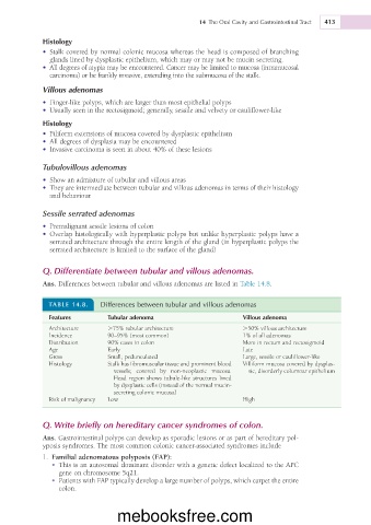
Q. Differentiate between tubular and villous adenomas.
Ans. Differences between tubular and villous adenomas are listed in Table 14.8.
TABLE 14.8. Differences between tubular and villous adenomas
Features Tubular adenoma Villous adenoma
Architecture .75% tubular architecture .50% villous architecture
Incidence 90–95% (most common) 1% of all adenomas
Distribution 90% cases in colon More in rectum and rectosigmoid
Age Early Late
Gross Small; pedunculated Large, sessile or cauliflower-like
Histology Stalk has fibromuscular tissue and prominent blood Villiform mucosa covered by dysplas-
vessels; covered by non-neoplastic mucosa. tic, disorderly columnar epithelium
Head region shows tubule-like structures lined
by dysplastic cells (instead of the normal mucin-
secreting colonic mucosa)
Risk of malignancy Low High
Q. Write briefly on hereditary cancer syndromes of colon.
Ans. Gastrointestinal polyps can develop as sporadic lesions or as part of hereditary pol-
yposis syndromes. The most common colonic cancer-associated syndromes include
1. Familial adenomatous polyposis (FAP):
• This is an autosomal dominant disorder with a genetic defect localized to the APC
gene on chromosome 5q21.
• Patients with FAP typically develop a large number of polyps, which carpet the entire
colon.
mebooksfree.com