
Page 431 - Concise Pathology for Exam Preparation ( PDFDrive )
P. 431
416
416 SECTION II Diseases of Organ Systems
Gross Morphology
Can be found anywhere in the colon and are typically seen as exophytic polypoid (right-side
colon) or annular constricting (left-side colon) growths
Microscopic Features
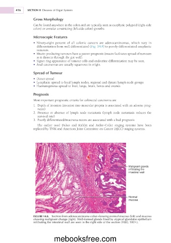
• Ninety-eight percent of all colonic cancers are adenocarcinomas, which vary in
differentiation from well differentiated (Fig. 14.8) to poorly differentiated anaplastic
tumours.
• Mucin-producing tumours have a poorer prognosis (mucin facilitates spread of tumours
as it dissects through the gut wall).
• Signet ring appearance of tumour cells and endocrine differentiation may be seen.
• Anal carcinomas are usually squamous in origin.
Spread of Tumour
• Direct spread
• Lymphatic spread to local lymph nodes, regional and distant lymph node groups
• Haematogenous spread to liver, lungs, brain, bones and ovaries
Prognosis
Most important prognostic criteria for colorectal carcinoma are
1. Depth of invasion (invasion into muscular propria is associated with an adverse prog-
nosis)
2. Presence or absence of lymph node metastasis (lymph node metastasis reduces the
survival rate)
3. Poorly differentiated/mucinous tutors are associated with a bad prognosis
The earlier used Dukes and Kirklin and Astler–Coller staging systems have been
replaced by TNM and American Joint Committee on Cancer (AJCC) staging systems.
Malignant glands
infiltrating the
intestinal wall
Normal
mucosa
FIGURE 14.8. Section from adenocarcinoma colon showing normal mucosa (left) and mucosa
showing malignant change (right). Well-formed glands lined by atypical glandular epithelium
infiltrating the intestinal wall are seen in the right side of the section (H&E; 1003).
mebooksfree.com