
Page 426 - Concise Pathology for Exam Preparation ( PDFDrive )
P. 426
14 The Oral Cavity and Gastrointestinal Tract 411
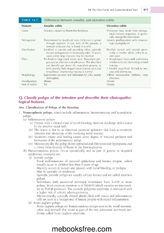
TABLE 14.7. Differences between amoebic and ulcerative colitis
Features Amoebic colitis Ulcerative colitis
Cause Infective; caused by Entamoeba histolytica Unknown; may result from dysregu-
lated immune responses, in geneti-
cally susceptible individuals
Pathogenesis Transmission by faecal-oral route (infection is spread Genetic predisposition with immuno-
through ingestion of cyst form of the parasite, a logic dysregulation
resistant structure that is found in stools)
Distribution Localized to caecum and ascending colon, sigmoid, Involves rectum and extends proxi-
rectum and appendix in decreasing order. In severe mally to involve whole colon in se-
cases, entire large intestine may be involved vere cases
Ulcer Pin-head to large-sized ulcers seen. Muscularis pro- • Broad-based ulcers with continuous
pria acts as a barrier to trophozoites. The ulcer fans involvement; no intervening normal
out laterally just above the muscularis propria, to mucosa.
form discrete flask-shaped ulcers (narrow neck and • Usually superficial: limited to mu-
broad base). Intervening mucosa is normal cosa and submucosa
Morphology Liquefactive necrosis; few inflammatory cells; mainly Diffuse mononuclear infiltrate, crypt
neutrophils abscesses
Pseudopolyps Absent Present
Risk of cancer Nil Present
Q. Classify polyps of the intestine and describe their clinicopatho-
logical features.
Ans. Classification of Polyps of the Intestine
1. Nonneoplastic polyps, which include inflammatory, hamartomatous and hyperplastic
polyps.
(a) Inflammatory polyps
(i) Present with a clinical triad of rectal bleeding, mucous discharge and a lesion
in the anterior rectal wall.
(ii) The lesion is due to an abnormal anorectal sphincter that leads to recurrent
abrasion and ulceration of the overlying rectal mucosa.
(iii) Recurrent injury and healing causes some degree of mucosal prolapse and
formation of the inflammatory polyp.
(iv) Microscopically, the polyp shows epithelial and fibromuscular hyperplasia and
a mixed inflammatory infiltrate in the lamina propria.
(b) Hamartomatous polyps: Occur sporadically and as part of genetic or acquired
syndromes, examples are:
(i) Juvenile polyps
- Focal malformations of mucosal epithelium and lamina propria, which
usually occur in children less than 5 years of age
- Majority occurs in rectum and present with rectal bleeding or prolapse
- May be sporadic or syndromic
- Sporadic juvenile polyps are usually solitary lesions and are called retention
polyps.
- Individuals with autosomal dominant inheritance have 3–100 or more
polyps. Most common mutation is of SMAD4 (which encodes an intermedi-
ate in TGF-b pathway). The juvenile polyposis syndrome is associated with
a higher risk of colonic adenocarcinoma.
- Microscopically, cystically dilated glands filled with mucin and inflammatory
cells are seen in a background of lamina propria with mixed inflammation.
(ii) Peutz–Jeghers polyps
- Peutz–Jeghers polyps are hamartomatous polyps seen in the small intestine,
colon and stomach that occur as part of the rare autosomal dominant syn-
drome called Peutz–Jeghers syndrome.
mebooksfree.com