
Page 425 - Concise Pathology for Exam Preparation ( PDFDrive )
P. 425
410
410 SECTION II Diseases of Organ Systems
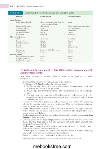
TABLE 14.6. Differences between Crohn disease and ulcerative colitis
Features Crohn disease Ulcerative colitis
Gross features
Bowel region affected Ileum, sometimes colon (may in- Colon
volve any part of GIT)
Pattern of distribution Skip lesions Diffuse, continuous involvement
Stricture formation Early Late, uncommon
Intestinal wall Thickened Thinned out
Intestinal dilatation Absent Present
Progression Antegrade Retrograde
Microscopic features
Ulcers Deep linear Superficial
Pseudo polyps Absent Present
Lymphoid reaction Marked Mild
Fibrosis Marked Mild
Serositis Marked Absent or mild
Granulomas Present in 50% of the cases Absent
Fistula/sinus Present Absent
Clinical features
Fat/vitamin malabsorption Present Absent
Malignant potential Less More
Response to surgery Poor Good
Q. Write briefly on amoebic colitis. Differentiate between amoebic
and ulcerative colitis.
Ans. Salient Features of Amoebic Colitis is caused by the protozoan Entamoeba
histolytica.
• Amoebic colitis is caused by the protozoan Entamoeba histolytica.
• The life cycle of E. histolytica has the following stages:
1. Trophozoite stage: Spherical to oval trophozoites can be demonstrated in the stool
of patients who exhibit acute symptoms.
2. Precyst stage: The trophozoite converts into a precyst form in the colon of the pa-
tient.
3. Cyst stage: Amoebic cysts have a thick chitinous wall and four nuclei. Infection
occurs by the faecal route due to ingestion of food contaminated with the faeces
containing the cysts.
• The cysts are resistant to gastric acid and are passed as it is to the colon where they
colonize the epithelial surface to release trophozoites. Most frequent location of coloni-
zation is caecum and ascending colon.
• Trophozoites produce a lytic substance which aids in invasion of the crypts. They then
burrow laterally into the lamina propria to form a superficial flask-shaped ulcer with a
narrow neck and wide base.
• Trophozoites may reach the liver by invading blood vessels to produce an amoebic liver
abscess in about 40% cases.
• Clinically patient in the acute stage presents with abdominal pain and bloody diar-
rhoea. Amoebic liver abscess typically manifests with right upper quadrant pain,
low-grade fever and weight loss.
• Trophozoites can microscopically be demonstrated in the surface of the ulcer which
shows both acute and chronic inflammation.
• Thickening of the intestinal wall with napkin ring–like constriction (ameboma) may
occasionally occur and can be confused with malignancy.
• Diagnosis is based on stool examination, serology and radiology.
Differences between amoebic and ulcerative colitis are listed in Table 14.7.
mebooksfree.com