
Page 457 - Concise Pathology for Exam Preparation ( PDFDrive )
P. 457
442 SECTION II Diseases of Organ Systems
Pathology
• Early lesions show dense lymphocytic and plasma cell infiltrate around small bile ducts
in the portal tracts.
• Late lesions show chronic granulomatous inflammation destroying the interlobular bile
ducts (florid duct lesion), resulting in fibrosis and later cirrhosis of the liver.
• In both early and late stages, there is marked hepatomegaly, contrary to other end-stage
liver diseases which show a small shrunken liver. This is probably due to the minimal
hepatocytic loss and extensive regeneration, typical of PBC.
PSC
Salient Features
• PSC is an immune-mediated chronic cholestatic disease characterized by progressive
concentric periductal (onion skin) fibrosis and destruction of extrahepatic and large
intrahepatic bile ducts. It has the following features:
• Median age is 30 years.
• Patient presents with fatigue, pruritis, jaundice, increased ALP levels and other features
of chronic cholestatic liver disease.
• Patchy involvement of the biliary tree results in characteristic ‘beading’ appearance of
the affected segment during a retrograde cholangiogram.
• Commonly coexists with inflammatory bowel disease, pancreatitis and retroperitoneal
fibrosis.
• Sixty-five percent patients are ANCA-positive.
• Cholangiocarcinomas may develop in 10–15% cases.
Pathology
• Obstruction of intrahepatic bile ducts leads to proliferation of bile ductules, inflamma-
tion and necrosis of adjacent periportal hepatic parenchyma and cholestasis.
• Large bile ducts show periductal fibrosis that obliterates the lumen leaving a solid cord-like
scar with a few inflammatory cells.
• Primary biliary cirrhosis and primary sclerosing cholangitis eventually lead to
end-stage liver disease (liver becomes hard and finely granular and shows yellow-
green pigmentation).
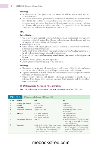
Q. Differentiate between PBC and PSC.
Ans. The differences between PBC and PSC are summarized in Table 15.4.
TABLE 15.4. Differences between PBC and PSC
S. No. Feature PBC PSC
1 Average age affected 50 years 30 years
2 Gender 90% females 70% females
3 Evolution Progressive Unpredictable
4 Associated conditions Sjögren syndrome Inflammatory bowel disease
Pancreatitis
5 Serology 95% AMA positivity 5% AMA positivity
50% ANA positivity 6% ANA positivity
40% ANCA positivity 65% ANCA positivity
6 Radiological features Normal Beaded appearance of the affected seg-
ment on a retrograde cholangio-
gram is diagnostic.
7 Pathology Small- and medium-sized intrahepatic Causes progressive sclerosing destruc-
bile ducts are affected. Large intrahe- tion of bile ducts of all sizes. Extra-
patic ducts and the extrahepatic bili- hepatic and large intrahepatic bile
ary structures are not involved. ducts are mainly involved.
mebooksfree.com