
Page 459 - Concise Pathology for Exam Preparation ( PDFDrive )
P. 459
444 SECTION II Diseases of Organ Systems
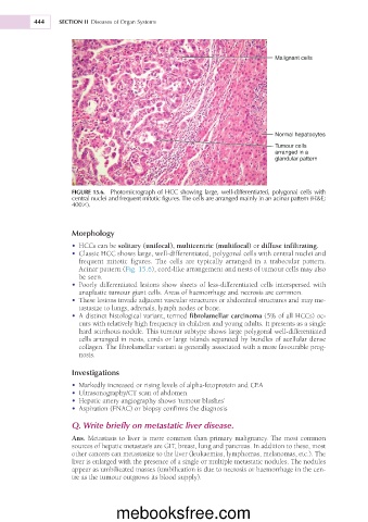
Malignant cells
Normal hepatocytes
Tumour cells
arranged in a
glandular pattern
FIGURE 15.6. Photomicrograph of HCC showing large, well-differentiated, polygonal cells with
central nuclei and frequent mitotic figures. The cells are arranged mainly in an acinar pattern (H&E;
4003).
Morphology
• HCCs can be solitary (unifocal), multicentric (multifocal) or diffuse infiltrating.
• Classic HCC shows large, well-differentiated, polygonal cells with central nuclei and
frequent mitotic figures. The cells are typically arranged in a trabecular pattern.
Acinar pattern (Fig. 15.6), cord-like arrangement and nests of tumour cells may also
be seen.
• Poorly differentiated lesions show sheets of less-differentiated cells interspersed with
anaplastic tumour giant cells. Areas of haemorrhage and necrosis are common.
• These lesions invade adjacent vascular structures or abdominal structures and may me-
tastasize to lungs, adrenals, lymph nodes or bone.
• A distinct histological variant, termed fibrolamellar carcinoma (5% of all HCCs) oc-
curs with relatively high frequency in children and young adults. It presents as a single
hard scirrhous nodule. This tumour subtype shows large polygonal well-differentiated
cells arranged in nests, cords or large islands separated by bundles of acellular dense
collagen. The fibrolamellar variant is generally associated with a more favourable prog-
nosis.
Investigations
• Markedly increased or rising levels of alpha-fetoprotein and CEA
• Ultrasonography/CT scan of abdomen
• Hepatic artery angiography shows ‘tumour blushes’
• Aspiration (FNAC) or biopsy confirms the diagnosis
Q. Write briefly on metastatic liver disease.
Ans. Metastasis to liver is more common than primary malignancy. The most common
sources of hepatic metastasis are GIT, breast, lung and pancreas. In addition to these, most
other cancers can metastasize to the liver (leukaemias, lymphomas, melanomas, etc.). The
liver is enlarged with the presence of a single or multiple metastatic nodules. The nodules
appear as umbilicated masses (umbilication is due to necrosis or haemorrhage in the cen-
tre as the tumour outgrows its blood supply).
mebooksfree.com