
Page 462 - Concise Pathology for Exam Preparation ( PDFDrive )
P. 462
15 Diseases of the Hepatobiliary System and Pancreas 447
• Stones are often present in the neck of the gallbladder or the cystic duct.
• Gallbladder lumen is filled with cloudy or turbid bile with or without admixed pus.
• When the contained exudate becomes pure pus, the condition is called empyema.
• In severe cases, gallbladder is transformed into a green-black necrotic organ (gan-
grenous cholecystitis).
• Histologically, the wall shows oedema, vascular congestion and neutrophilic infiltrate.
2. Chronic cholecystitis
Salient features:
• May follow repeated attacks of acute cholecystitis or develop without any history of
previous attacks.
• Clinically, it presents with recurrent attacks of colicky epigastric or right upper quad-
rant pain, nausea, vomiting and intolerance to fatty food.
• Usually associated with gallstones in the lumen or presence of biliary gravel (thick
viscous bile with micro-concretions).
• Chronic acalculus cholecystitis causes symptoms and morphological alterations
similar to chronic calculus cholecystitis.
Pathology:
• Serosa is dull and opaque and may show adhesions.
• Mucosa is oedematous, focally ulcerated or indurated.
• Gallbladder may be contracted, of normal size, or enlarged.
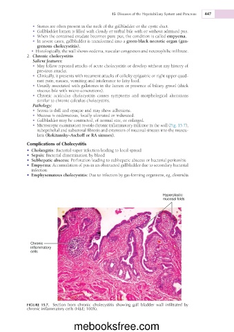
• Microscopic examination reveals chronic inflammatory infiltrate in the wall (Fig. 15.7),
subepithelial and subserosal fibrosis and extension of mucosal sinuses into the muscu-
laris (Rokitansky–Aschoff or RA sinuses).
Complications of Cholecystitis
• Cholangitis: Bacterial super infection leading to local spread
• Sepsis: Bacterial dissemination by blood
• Subhepatic abscess: Perforation leading to subhepatic abscess or bacterial peritonitis
• Empyema: Accumulation of pus in an obstructed gallbladder due to secondary bacterial
infection
• Emphysematous cholecystitis: Due to infection by gas-forming organisms, eg, clostridia
Hyperplastic
mucosal folds
Chronic
inflammatory
cells
FIGURE 15.7. Section from chronic cholecystitis showing gall bladder wall infiltrated by
chronic inflammatory cells (H&E; 100X).
mebooksfree.com