
Page 461 - Concise Pathology for Exam Preparation ( PDFDrive )
P. 461
446 SECTION II Diseases of Organ Systems
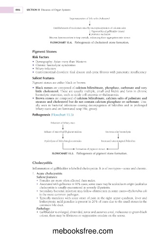
Supersaturation of bile with cholesterol
Establishment of nucleation sites by microprecipitation of calcium salts
Hypomotility of gallbladder (stasis)
promotes nucleation
Mucous hypersecretion to trap crystals, enhancing their aggregation into stones
FLOWCHART 15.4. Pathogenesis of cholesterol stone formation.
Pigment Stones
Risk Factors
• Demography: Asian more than Western
• Chronic haemolytic syndromes
• Biliary infection
• Gastrointestinal disorders: Ileal disease and cystic fibrosis with pancreatic insufficiency
Salient Features
Pigment stones are either black or brown:
• Black stones are composed of calcium bilirubinate, phosphate, carbonate and very
little cholesterol. These are usually multiple, small and friable and form in chronic
haemolytic anaemias, such as sickle cell anaemia or thalassaemia.
• Brown stones are composed of calcium bilirubinate, calcium salts of palmitate and
stearate and cholesterol but do not contain calcium phosphate or carbonate. Usu-
ally seen in bacterial infections causing deconjugation of bilirubin and in prolonged
biliary stasis and are laminated soap like, greasy.
Pathogenesis (Flowchart 15.5)
Infection of biliary tract
Release of microbial β-glucuronidase Intravascular haemolysis
Hydrolysis of bilirubin glucuronides Increased unconjugated bilirubin
Formation of pigment stones
FLOWCHART 15.5. Pathogenesis of pigment stone formation.
Cholecystitis
Inflammation of gallbladder is labelled cholecystitis. It is of two types—acute and chronic.
1. Acute cholecystitis
Salient features:
• Females are more often affected than males.
• Associated with gallstones in 90% cases; some cases may be acalculus in origin (acalculus
cholecystitis is usually encountered in severely ill patients.
• Secondary bacterial infection may follow obstruction in some cases—Escherichia coli
is the most common pathogen.
• Typically manifests with acute onset of pain in the right upper quadrant, fever and
leukocytosis; mild jaundice is present in 20% of cases due to the small stones in the
common bile duct.
Pathology:
• Gallbladder is enlarged, distended, tense and assumes a red, violaceous to green-black
colour, there may be fibrinous or suppurative exudate on the serosa.
mebooksfree.com