
Page 505 - Concise Pathology for Exam Preparation ( PDFDrive )
P. 505
490 SECTION II Diseases of Organ Systems
• An undescended testis is called retractile when it can be manipulated into scrotum
where it remains without tension. On the other hand, when it can be manipulated into
upper scrotum but retracts when released, it is called gliding.
• Cryptorchidism is unilateral in about 80% cases and bilateral in the remaining. Most
cases are clinically asymptomatic and discovered only on physical examination.
• Cryptorchid testes can be brought into the scrotum by a surgical procedure called an
‘orchiopexy’.
• Untreated cases may be associated with reduced fertility, increased risk of testicular
germ cell tumours and are also more prone to torsion, infarction and inguinal hernia.
• On gross examination, the cryptorchid testis is small and fibrotic. Histologically, there
is marked reduction in the number of germ cells.
Q. Write briefly on testicular atrophy.
Ans. Testicular atrophy is a regressive change which can have a varied aetiology.
Causes
• Progressive atherosclerotic narrowing of testicular blood vessels, as in old age
• End stage of all inflammatory conditions (orchitis)
• Cryptorchidism
• Hypopituitarism
• Obstruction of flow to semen
• Malnutrition and cachexia
• Prolonged administration of female sex hormones
• Exhaustion atrophy due to high level of pituitary follicle-stimulating hormone
• Klinefelter syndrome
Gross Morphology
Testes are small in size and firm in consistency due to fibrotic changes.
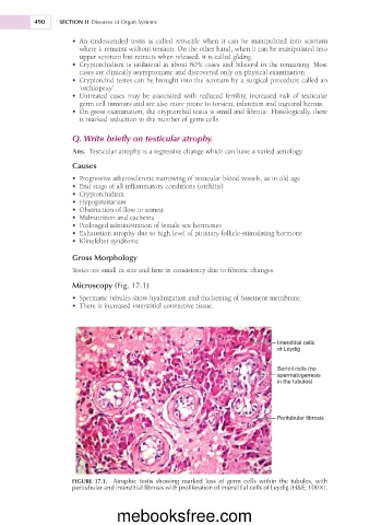
Microscopy (Fig. 17.1)
• Spermatic tubules show hyalinization and thickening of basement membrane.
• There is increased interstitial connective tissue.
Interstitial cells
of Leydig
Sertoli cells (no
spermatogenesis
in the tubules)
Peritubular fibrosis
FIGURE 17.1. Atrophic testis showing marked loss of germ cells within the tubules, with
peritubular and interstitial fibrosis with proliferation of interstitial cells of Leydig (H&E; 1003).
mebooksfree.com