
Page 507 - Concise Pathology for Exam Preparation ( PDFDrive )
P. 507
492 SECTION II Diseases of Organ Systems
- Stage II: Spread confined to retroperitoneal lymph nodes below the diaphragm
- Stage III: Metastasis outside the retroperitoneal lymph nodes or above the
diaphragm
Note: Most seminomas present in Stage I disease; lymph nodes are commonly involved;
haematogenous spread is a late manifestation. Most NSGCTs present in Stage II or III
disease; haematogenous spread is an early manifestation.
1. Germ cell tumours
(a) Seminomatous germ cell tumours (SGCTs)
(i) Typical/classical seminoma (85%)
Clinical features:
• Most common type of germ cell tumour
• Peak age: third decade; never seen in infants
• Extremely radiosensitive
�
Gross morphology:
• Classical seminomas are large tumours which may replace the entire testis but
the testicular shape is maintained.
• Cut surface is homogeneous, grey-white and lobulated.
• Haemorrhage and necrosis are rare.
• Tunica albuginea is generally intact; however, occasional extension to epididymis,
spermatic cord and scrotal sac may be seen.
�
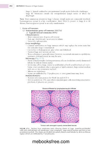
Microscopy (Fig. 17.2):
�
• Sheets of monomorphic-looking seminoma cells are divided into poorly demarcated
lobules by delicate fibrous septae.
• Seminoma cell is a large, round-to-polyhedral cell with a well-defined cell mem-
brane; clear cytoplasm (due to glycogen or lipid contents), large central nucleus
with one or two prominent nucleoli.
• Mitoses are infrequent.
• Septae are infiltrated by T lymphocytes; at times granulomas may form.
Immunochemistry:
• Tumour cells stain positive for PLAP, kit and OCT 4.
• HCG is positive in 15% cases where syncytial giant cells resembling syncytiotro-
phoblasts of placenta are present.
FIGURE 17.2. Section from seminoma testis showing sheets of large, round-to-polyhedral
cells with well-defined cell membrane; clear cytoplasm, large central nucleus and one or two
prominent nucleoli. The sheets are divided into poorly demarcated lobules by delicate fibrous
septae which are infiltrated by T lymphocytes.
mebooksfree.com