
Page 518 - Concise Pathology for Exam Preparation ( PDFDrive )
P. 518
18 Female Genital System 503
Fallopian Tube
• Average size is 10 cm 3 1 mm; it is divided into four anatomical regions, namely, isthmus,
ampulla, fimbriae and abdominal opening.
• Mucosa is lined by three cell types—ciliated columnar, nonciliated columnar and
intercalated cells (inactive secretory cells).
• Anterior to the tubes there is insertion of round ligament. Most lateral portion of broad
ligament is called infundibulopelvic or suspensory ligament and transmits ovarian
vessels and nerves.
Nulliparous Uterus
• Weighs 30–40 g, with an average size of 7.5 cm 3 5 cm 3 2.5 cm.
• It has an inverted flattened pear appearance with the presence of a constriction called
isthmus.
• Anterior peritoneal reflection is at isthmus; posteriorly peritoneum covers the entire
uterus and passes down to cover the upper portion of the vagina.
• Vaginal portion is covered by moist, smooth vaginal epithelium.
• Cervical canal is narrow and fusiform.
Multiparous Uterus
Weighs 60–70 g, with an average size of 10 cm 3 5 cm 3 4 cm.
Postmenopausal Uterus
• Atrophic/more fibrous
• Cervix less prominent
• Uterus is anteflexed (sharply bent forward upon vagina)
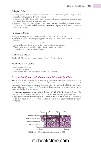
Q. Write briefly on cervical intraepithelial neoplasia (CIN).
Ans. CIN is a precancerous lesion frequently associated with HPV infection. HPV is a
sexually transmitted DNA virus which can lead to CIN as well as invasive squamous cell
carcinoma. The spectrum of changes associated with progression of CIN to invasive carci-
noma is illustrated in Figure 18.1. According to Bethesda system, precancerous lesions of
cervix are divided into two groups:
• Low-grade squamous intraepithelial lesion or LSIL (CIN I): Associated with HPV
types 6, 11, 42 and 44 (also known as HPV types with low oncogenic potential).
• High-grade squamous intraepithelial lesion or HSIL (CIN II and III): Associated
with HPV types 16, 18, 31, 33 and 45 (also known as HPV types with high oncogenic
potential).
Normal CIN1 CIN2 CIN3
Stratum corneum
Stratum granulosum
Stratum spinosum
Stratum basale
Basement
membrane Stroma
FIGURE 18.1. Histopathological spectrum of CIN.
mebooksfree.com