
Page 521 - Concise Pathology for Exam Preparation ( PDFDrive )
P. 521
506 SECTION II Diseases of Organ Systems
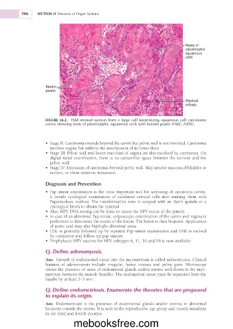
Nests of
pleomorphic
squamous
cells
Keratin
pearls
Atypical
mitosis
FIGURE 18.2. H&E-stained section from a large cell keratinizing squamous cell carcinoma
cervix showing nests of pleomorphic squamous cells with keratin pearls (H&E; 200X).
• Stage II: Carcinoma extends beyond the cervix but pelvic wall is not involved. Carcinoma
involves vagina but without the involvement of its lower third.
• Stage III: Pelvic wall and lower one-third of vagina are also involved by carcinoma. On
digital rectal examination, there is no cancer-free space between the tumour and the
pelvic wall.
• Stage IV: Extension of carcinoma beyond pelvic wall. May involve mucosa of bladder or
rectum, or show systemic metastasis.
Diagnosis and Prevention
• Pap smear examination is the most important tool for screening of carcinoma cervix.
It entails cytological examination of exfoliated cervical cells after staining them with
Papanicolaou method. The transformation zone is scraped with an Ayer’s spatula or a
cytological brush to obtain the material.
• Also, HPV DNA testing can be done to assess the HPV status of the patient.
• In case of an abnormal Pap smear, colposcopic examination of the cervix and vagina is
performed to determine the extent of the lesion. The lesion is then biopsied. Application
of acetic acid may also highlight abnormal areas.
• LSIL is generally followed up by repeated Pap smear examination and HSIL is excised
by conization and follow-up pap smears.
• Prophylactic HPV vaccine for HPV subtypes 6, 11, 16 and 18 is now available.
Q. Define adenomyosis.
Ans. Growth of endometrial tissue into the myometrium is called adenomyosis. Clinical
features of adenomyosis include irregular, heavy menses and pelvic pain. Microscopy
shows the presence of nests of endometrial glands and/or stroma well down in the myo-
metrium between the muscle bundles. The endometrial tissue must be separated from the
basalis by at least 2–3 mm.
Q. Define endometriosis. Enumerate the theories that are proposed
to explain its origin.
Ans. Endometriosis is the presence of endometrial glands and/or stroma in abnormal
locations outside the uterus. It is seen in the reproductive age group and mostly manifests
in the third and fourth decades.
mebooksfree.com