
Page 520 - Concise Pathology for Exam Preparation ( PDFDrive )
P. 520
18 Female Genital System 505
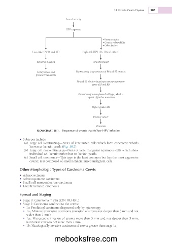
Sexual activity
HPV exposure
• Immune status
• Genetic vulnerability
• Other factors
Low-risk HPV (6 and 11) High-risk HPV (16, 18 and others)
Episomal infection Viral integration
Condylomata and Expression of large amounts of E6 and E7 proteins
precancerous lesions
E6 and E7 block or inactivate tumour suppressor
genes p53 and RB
Formation of a transformed cell type, which is
capable of further mutations
Higher grade CIN
Invasive cancer
Metastasis
FLOWCHART 18.1. Sequence of events that follow HPV infection.
• Subtypes include
(a) Large cell keratinizing—Nests of keratinized cells which form concentric whorls
known as keratin pearls (Fig. 18.2).
(b) Large cell nonkeratinizing—Nests of large malignant squamous cells which show
individual cell keratinization but no keratin pearls.
(c) Small cell carcinoma—This type is the least common but has the most aggressive
course; it is composed of small nonkeratinized malignant cells.
Other Morphologic Types of Carcinoma Cervix
• Adenocarcinoma
• Adenosquamous carcinoma
• Small cell neuroendocrine carcinoma
• Undifferentiated carcinoma
Spread and Staging
• Stage 0: Carcinoma in situ (CIN III, HSIL)
• Stage I: Carcinoma confined to the cervix:
• 1a: Preclinical carcinoma diagnosed only by microscopy
• 1a 1 : Minimally invasive carcinoma (invasion of stroma not deeper than 3 mm and not
wider than 7 mm)
• 1a 2 : Microscopic invasion of stroma more than 3 mm and not deeper than 5 mm;
horizontal invasion not more than 7 mm
• 1b: Histologically invasive carcinoma of cervix greater than stage 1a 2
mebooksfree.com