
Page 524 - Concise Pathology for Exam Preparation ( PDFDrive )
P. 524
18 Female Genital System 509
Atypical Hyperplasia
Complex architectural patterns with cellular atypia is the hallmark. Atypical hyperplasia is
difficult to differentiate from a well-differentiated carcinoma on biopsy. About 20–30% of
these cases show foci of endometrial carcinoma on hysterectomy.
Q. Write in detail on the aetiology, clinical features and morphology
of endometrial carcinoma.
Ans. Endometrial carcinoma is the most common cancer of female genital tract. It presents
with irregular or postmenopausal bleeding and leucorrhoea.
Types (Table 18.1)
• Type 1 (constitutes 80% of all cases; is oestrogen associated).
• Type 2 (less common; not associated with hyperoestrogenaemia).
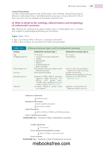
TABLE 18.1. Differences between Types I and II of endometrial carcinoma
Features Endometrial carcinoma Type 1 Endometrial carcinoma Type II
Age 55–60 years 65–75 years
Predisposing factors • Unopposed oestrogen stimulation Thin physique
• Obesity
• Hypertension
• Diabetes
• Nulliparity/infertility
• Breast carcinoma
Morphological type Endometrioid carcinoma (mimics normal Serous or clear cell type (mimics
endometrial glands) subtypes of ovarian carcinoma)
Precursor Endometrial hyperplasia Atrophic endometrium
Endometrial intraepithelial carcinoma
Molecular genetics Mutations in PTEN, ARID1A (chromatin Mutations in P53 and PIK3CA
regulator), KRAS, b-catenin, p53, (Flowchart 18.3)
PIK3CA, FGF2 (growth factor),
CTNNB1 (Wnt signalling) and micro-
satellite instability (Flowchart 18.2)
Outcome Low-grade malignancy; spreads mainly Aggressive; intraperitoneal and
via lymphatics lymphatic spread is common
Proliferative endometrium
PTEN abnormality
Hyperplasia without atypia
MLH1 and KRAS abnormalities
Microsatellite instability
Atypical hyperplasia
ARID 1A, PIK3CA, CTNNB1 and FGFR2 abnormalities
Grade 1 endometrioid carcinoma
FLOWCHART 18.2. Evolution of Type I endometrial carcinoma.
Atrophic endometrium
TP53 mutation
Serous endometrial intraepithelial carcinoma
FBXW7, PPP2RIA, CCNE1 abnormalities
Serous carcinoma
FLOWCHART 18.3. Evolution of Type II endometrial carcinoma.
mebooksfree.com