
Page 529 - Concise Pathology for Exam Preparation ( PDFDrive )
P. 529
514 SECTION II Diseases of Organ Systems
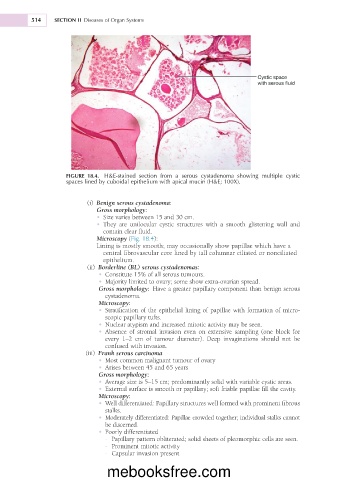
Cystic space
with serous fluid
FIGURE 18.4. H&E-stained section from a serous cystadenoma showing multiple cystic
spaces lined by cuboidal epithelium with apical mucin (H&E; 100X).
(i) Benign serous cystadenoma:
Gross morphology:
• Size varies between 15 and 30 cm.
• They are unilocular cystic structures with a smooth glistening wall and
contain clear fluid.
Microscopy (Fig. 18.4):
Lining is mostly smooth; may occasionally show papillae which have a
central fibrovascular core lined by tall columnar ciliated or nonciliated
epithelium.
(ii) Borderline (BL) serous cystadenomas:
• Constitute 15% of all serous tumours.
• Majority limited to ovary; some show extra-ovarian spread.
Gross morphology: Have a greater papillary component than benign serous
cystadenoma.
Microscopy:
• Stratification of the epithelial lining of papillae with formation of micro-
scopic papillary tufts.
• Nuclear atypism and increased mitotic activity may be seen.
• Absence of stromal invasion even on extensive sampling (one block for
every 1–2 cm of tumour diameter). Deep invaginations should not be
confused with invasion.
(iii) Frank serous carcinoma
• Most common malignant tumour of ovary
• Arises between 45 and 65 years
Gross morphology:
• Average size is 5–15 cm; predominantly solid with variable cystic areas.
• External surface is smooth or papillary; soft friable papillae fill the cavity.
Microscopy:
• Well differentiated: Papillary structures well formed with prominent fibrous
stalks.
• Moderately differentiated: Papillae crowded together; individual stalks cannot
be discerned.
• Poorly differentiated
- Papillary pattern obliterated; solid sheets of pleomorphic cells are seen.
- Prominent mitotic activity
- Capsular invasion present
mebooksfree.com