
Page 545 - Concise Pathology for Exam Preparation ( PDFDrive )
P. 545
530 SECTION II Diseases of Organ Systems
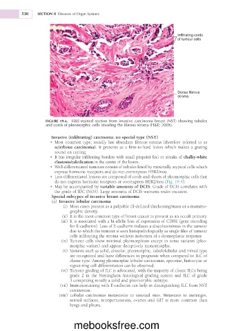
Infiltrating cords
of tumour cells
Dense fibrous
stroma
FIGURE 19.4. H&E-stained section from invasive carcinoma breast (NST) showing tubules
and cords of pleomorphic cells invading the fibrous stroma (H&E; 200X).
Invasive (infiltrating) carcinoma; no special type (NST)
• Most common type; usually has abundant fibrous stroma (therefore referred to as
scirrhous carcinoma). It presents as a firm-to-hard lesion which makes a grating
sound on cutting.
• It has irregular infiltrating borders with small pinpoint foci or streaks of chalky-white
elastosis/calcification in the centre of the lesion.
• Well-differentiated tumours consist of tubules lined by minimally atypical cells which
express hormone receptors and do not overexpress HER2/neu.
• Less-differentiated lesions are composed of cords and sheets of pleomorphic cells that
do not express hormone receptors or overexpress HER2/neu (Fig. 19.4).
• May be accompanied by variable amounts of DCIS. Grade of DCIS correlates with
the grade of IDC (NOS). Large amounts of DCIS warrants wider excision.
Special subtypes of invasive breast carcinoma
(a) Invasive lobular carcinoma
(i) Most cases present as a palpable ill-defined thickening/mass or a mammo-
graphic density.
(ii) It is the most common type of breast cancer to present as an occult primary.
(iii) It is associated with a bi-allelic loss of expression of CDH1 (gene encoding
for E-cadherin). Loss of E-cadherin induces a discohesiveness in the tumour
due to which the tumour is seen histopathologically as single files of tumour
cells infiltrating the stroma without induction of a desmoplastic response
(iv) Tumour cells show minimal pleomorphism except in some variants (pleo-
morphic variant) and appear deceptively monomorphic.
(v) Variants such as solid, alveolar, pleomorphic, tubulolobular and mixed type
are recognized and have differences in prognosis when compared to ILC of
classic type. Among pleomorphic lobular carcinomas, apocrine, histiocytic or
signet-ring cell differentiation can be observed.
(vi) Tumour grading of ILC is advocated, with the majority of classic ILCs being
grade 2 in the Nottingham histological grading system and ILC of grade
3 comprising mostly a solid and pleomorphic subtype.
(vii) Immunostaining with E-cadherin can help in distinguishing ILC from NST
carcinomas.
(viii) Lobular carcinomas metastasize to unusual sites. Metastasis to meninges,
serosal surfaces, retroperitoneum, ovaries and GIT is more common than
lungs and pleura.
mebooksfree.com