
Page 540 - Concise Pathology for Exam Preparation ( PDFDrive )
P. 540
19 The Breast 525
Capsule
Slit-like ducts
Stromal tissue
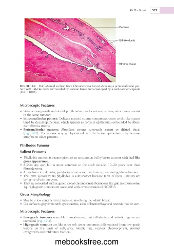
FIGURE 19.2. H&E-stained section from fibroadenoma breast showing a pericanalicular pat-
tern with slit-like ducts surrounded by stromal tissue and enveloped by a well-formed capsule
(H&E; 100X).
Microscopic Features
• Stromal overgrowth and ductal proliferation produces two patterns, which may coexist
in the same tumour:
• Intracanalicular pattern: Delicate myxoid stroma compresses ducts to slit-like spaces
lined by ductal epithelium, which appears as cords of epithelium surrounded by abun-
dant fibrous stroma.
• Pericanalicular pattern: Abundant stroma surrounds patent or dilated ducts
(Fig. 19.2). The stroma may get hyalinized and the lining epithelium may become
atrophic in older patients.
Phyllodes Tumour
Salient Features
• ‘Phyllodes tumour’ is a name given to an uncommon bulky breast tumour with leaf-like
gross appearance.
• Affects any age, but is more common in the sixth decade, 10–20 years later than
fibroadenoma.
• Arises from intralobular, periductal stroma and not from a pre-existing fibroadenoma.
• The term ‘cystosarcoma phyllodes’ is a misnomer because most of these tumours are
benign and without cysts.
• They are associated with acquired clonal chromosomal aberrations like gain in chromosome
1q. High-grade tumours are associated with overexpression of HOXB13.
Gross Morphology
• May be a few centimetres to massive, involving the whole breast.
• Cut surface is grey-white with cystic cavities, areas of haemorrhage and necrosis may be seen.
Microscopic Features
• Low-grade tumours resemble fibroadenoma, but cellularity and mitotic figures are
increased (Fig. 19.3).
• High-grade tumours are like other soft tissue sarcomas; differentiated from low-grade
lesions on the basis of cellularity, mitotic rate, nuclear pleomorphism, stromal
overgrowth and infiltrative borders.
mebooksfree.com