
Page 574 - Concise Pathology for Exam Preparation ( PDFDrive )
P. 574
20 Endocrinology 559
Two factors, namely ‘glucose-dependent insulinotropic polypeptide or GIP’ secreted by
endocrine k cells located in the small bowel and ‘glucagon-like peptide-1 or GLP-1Z’
secreted by L cells located in distal ileum and colon are released immediately after
food intake. They are called incretins and their stimulatory effect on secretion of in-
sulin from b cells is labelled ‘incretin effect’. This effect is blunted in Type II diabetes.
2. Signalling pathway
(a) Insulin receptor is a tetrameric protein composed of two a and two b chains.
(b) The b subunit cytosolic domain possesses tyrosine kinase activity.
(c) Insulin binds to the extracellular domain of a subunit to activate the b subunit
tyrosine kinase, leading to autophosphorylation of the receptor and phosphoryla-
tion of several intracellular substrate proteins, eg, family of insulin receptor sub-
strate proteins (IRS) proteins, which includes IRS1-4 and GAB1.
(d) The substrate proteins activate multiple downstream signal cascades including PI-
3k and MAP kinase pathways, which mediate the several actions of insulin.
(e) Insulin aids in the docking of glucose transporter unit GLUT-4 to the plasma mem-
brane (GLUT-4 promotes glucose uptake).
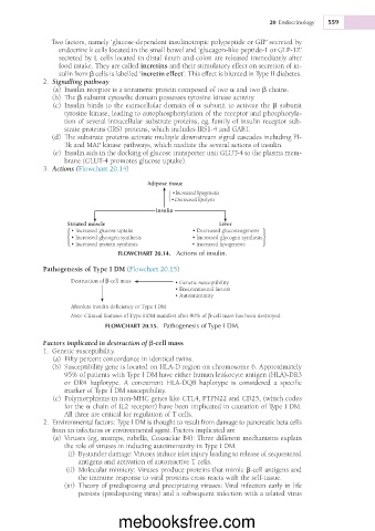
3. Actions (Flowchart 20.14)
Adipose tissue
• Increased lipogenesis
• Decreased lipolysis
Insulin
Striated muscle Liver
• Increased glucose uptake • Decreased gluconeogenesis
• Increased glycogen synthesis • Increased glycogen synthesis
• Increased protein synthesis • Increased lipogenesis
FLOWCHART 20.14. Actions of insulin.
Pathogenesis of Type I DM (Flowchart 20.15)
Destruction of β-cell mass • Genetic susceptibility
• Environmental factors
• Autoimmunity
Absolute insulin deficiency or Type I DM
Note: Clinical features of Type I DM manifest after 80% of β-cell mass has been destroyed.
FLOWCHART 20.15. Pathogenesis of Type I DM.
Factors implicated in destruction of b-cell mass
1. Genetic susceptibility.
(a) Fifty percent concordance in identical twins.
(b) Susceptibility gene is located on HLA-D region on chromosome 6. Approximately
95% of patients with Type I DM have either human leukocyte antigen (HLA)-DR3
or DR4 haplotype. A concurrent HLA-DQ8 haplotype is considered a specific
marker of Type I DM susceptibility.
(c) Polymorphisms in non-MHC genes like CTL4, PTPN22 and CD25, (which codes
for the a chain of IL2 receptor) have been implicated in causation of Type I DM.
All three are critical for regulation of T cells.
2. Environmental factors: Type I DM is thought to result from damage to pancreatic beta cells
from an infectious or environmental agent. Factors implicated are
(a) Viruses (eg, mumps, rubella, Coxsackie B4): Three different mechanisms explain
the role of viruses in inducing autoimmunity in Type I DM.
(i) Bystander damage: Viruses induce islet injury leading to release of sequestered
antigens and activation of autoreactive T cells.
(ii) Molecular mimicry: Viruses produce proteins that mimic b-cell antigens and
the immune response to viral proteins cross reacts with the self-tissue.
(iii) Theory of predisposing and precipitating viruses: Viral infection early in life
persists (predisposing virus) and a subsequent infection with a related virus
mebooksfree.com