
Page 575 - Concise Pathology for Exam Preparation ( PDFDrive )
P. 575
560 SECTION II Diseases of Organ Systems
(precipitating virus) that shares antigenic epitopes, leads to an immune re-
sponse against the infected islet cells.
(b) Toxic chemicals
(c) Exposure to cow’s milk in infancy
(d) Cytotoxins
(e) Recent evidence suggests a role for vitamin D in the pathogenesis and prevention
of diabetes mellitus.
3. Autoimmune factors: Currently, autoimmunity is considered the major factor in the
pathophysiology of Type I DM. Evidence implicating autoimmunity includes
(a) Circulating islet cell (glutamic acid decarboxylase or GAD and antiinsulin) anti-
bodies
(b) b cells damage by cytokines (g IFN, TNF and IL1)
(c) Prominent insulitis (including cellular necrosis and lymphocytic infiltration)
(d) Tissue injury caused by macrophages activated by CD4 T cells
1
1
(e) Direct killing of b cells by CD8 T cells
(f) Increased prevalence of Type I DM in patients with other autoimmune diseases,
such as Graves disease, Hashimoto thyroiditis and Addison disease.
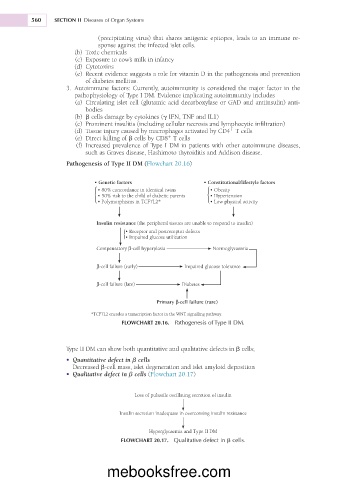
Pathogenesis of Type II DM (Flowchart 20.16)
• Genetic factors • Constitutional/lifestyle factors
• 80% concordance in identical twins • Obesity
• 50% risk to the child of diabetic parents • Hypertension
• Polymorphisms in TCF7L2* • Low physical activity
Insulin resistance (the peripheral tissues are unable to respond to insulin)
• Receptor and postreceptor defects
• Impaired glucose utilization
Compensatory β-cell hyperplasia Normoglycaemia
β-cell failure (early) Impaired glucose tolerance
β-cell failure (late) Diabetes
Primary β-cell failure (rare)
*TCF7L2 encodes a transcription factor in the WNT signalling pathway.
FLOWCHART 20.16. Pathogenesis of Type II DM.
Type II DM can show both quantitative and qualitative defects in b cells;
• Quantitative defect in b cells
Decreased b-cell mass, islet degeneration and islet amyloid deposition
• Qualitative defect in b cells (Flowchart 20.17)
Loss of pulsatile oscillating secretion of insulin
Insulin secretion inadequate in overcoming insulin resistance
Hyperglycaemia and Type II DM
FLOWCHART 20.17. Qualitative defect in b cells.
mebooksfree.com