
Page 1074 - Hematology_ Basic Principles and Practice ( PDFDrive )
P. 1074
Chapter 60 Myelodysplastic Syndromes 957
Fig. 60.3 GRANULOCYTIC DYSPLASIA. Granulocytic dysplasia is most evident in mature neutrophils
and can be contrasted to features of normal forms (far left, top), which are usually still present as a subpopula-
tion of the total cells in most cases. Granulocytic dysplasia is characterized by (left to right, starting at second
column) reduced cytoplasmic granulation, nuclear hypolobation (resulting in the binuclear or single-lobed
pseudo–Pelger-Huët forms), hypersegmentation, ringed forms (“rodent cells”), cells with nuclear twinning,
and cells with excessive nuclear excrescences. Photomicrographs are from a number of cases of refractory
cytopenia with multilineage dysplasia and refractory anemia with excess blasts.
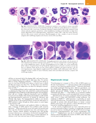
Fig. 60.4 MEGAKARYOCYTIC DYSPLASIA. Dysmegakaryopoiesis is most obvious with the presence of
micromegakaryocytes and abnormal larger forms (panels 2–5). These are compared with a normal megakaryo-
cyte at same magnification (panel 1, far left). Micromegakaryocytes have single, two, or four small nuclei,
which indicate a low-ploidy level. Normal low-ploidy megakaryocytes can be seen in the bone marrow, but
these are immature forms and do not have mature granular cytoplasm with platelet material, as do the
micromegakaryocytes. Larger dysplastic megakaryocytes have multiple, small, widely spaced nuclei. Photomi-
crographs are from a number of cases of refractory anemia with excess blasts and refractory cytopenia with
multilineage dysplasia.
still have an increased risk of developing AML, and some have pro-
posed designating any MDS patient with more than 2% marrow Megakaryocytic Lineage
310
blasts as having “oligoblastic leukemia.” ALIP has been shown to
occur in most cases of RAEB and in about a third of cases in which Thrombocytopenia is present in 25% to 50% of MDS patients at
311
the blast percentage is less than 5%. In these latter cases ALIP has the time of presentation, 296,306 and some patients with normal platelet
314
been shown to be an independent risk factor for subsequent develop- counts can have functional platelet defects. Laboratory abnormali-
ment of AML. 312 ties include prolonged bleeding times, defective granulation, and
In the peripheral blood, visible morphologic abnormalities include abnormal platelet aggregation indices mediated by hypofunctional
so-called pseudo Pelger-Huët cells, which have condensed chromatin platelet glycoprotein IIb/IIIa, leading to what is known as a
and bilobed nuclei resembling an old-fashioned pince-nez (true Pelger- Glanzmann-type defect. 315,316 These defects can occasionally manifest
Huët cells are a benign congenital abnormality seen in children). as spontaneous bleeding, or can be unmasked after trauma or surgery.
Granulocytes may display other nuclear abnormalities as well, includ- Thrombocytosis, by contrast, is relatively unusual in patients with
ing hypersegmentation reminiscent of vitamin B 12 deficiency, and MDS, except in those with MDS/MPN overlap syndromes, 5q−
317
aberrant ring shapes. The hypogranulation visible in the marrow syndrome, or RARS-T. JAK2 mutations, in particular, are associ-
typically persists in the peripheral blood, and the left shift typical of ated with thrombocytosis. Thrombocytosis can occasionally be subtle,
MDS marrows is often, though not always, present to some degree especially in patients with advanced disease, in whom a normal
in the blood as well. platelet count may in fact represent a relative thrombocytosis coun-
Despite these quantitative and qualitative defects in leukocytes, teracted by dwindling megakaryocytic reserves.
only a minority of MDS patients have problems with recurrent In the marrow, megakaryocytes are most commonly present in
313
infections. When infections do occur, they tend to be bacterial and normal or increased numbers. A number of morphologic abnormali-
often arise from the lower respiratory tract, skin, and mucous mem- ties can be observed, including abnormally small forms (micromega-
branes. Patients without absolute neutropenia may still develop karyocytes), hypersegmentation, and nuclear hypolobation (Fig.
recurrent infections as a consequence of abnormal neutrophil func- 60.4). 318,319 Megakaryocytes may also be abnormally distributed in
tion. Even though only some patients have recurrent infections, an clusters scattered throughout the marrow in MDS, rather than their
infection is the most common cause of MDS-associated death. normal parasinusoidal positioning.