
Page 1077 - Hematology_ Basic Principles and Practice ( PDFDrive )
P. 1077
960 Part VII Hematologic Malignancies
before the introduction of the IPSS-R, but subsequent studies have typically remain transfusion dependent, and management of iron
suggested that the WPSS and the IPSS-R are about equal in their overload then becomes increasingly important.
accuracy of predicting prognosis. 342 The designation of 5q− syndrome applies only to patients in
whom loss of 5q is the only cytogenetic abnormality. Patients in
whom 5q− is only one of several cytogenetic aberrations in fact tend
Specific Clinical Syndromes to have a worse prognosis than average, with a more rapid progression
to AML. As referenced previously, loss of 5q often occurs in tandem
Although MDS is a heterogeneous disorder, it contains a number of with TP53 mutations (or loss of 17p), another poor-prognosis
specific entities, either defined by cytogenetics, pathologic findings, combination. 149
or clinical features, which possess distinctive clinical features or bio-
logic behaviors. Some of these are discussed in more detail later.
Hypocellular Syndromes
The 5q− Syndrome Most patients with MDS have hypercellular or normocellular
marrows. Hypocellular marrows, in contrast, are found in fewer
Patients with isolated loss of the long arm of chromosome 5 [so-called than 15% of patients, and delineate the entity of hypoplastic MDS
224
del(5q) or 5q minus syndrome] are a unique group whose biology is (Fig. 60.5D–E). These patients have substantial overlap, both
described in more detail earlier. Clinically, 5q− syndrome is character- morphologically and clinically, with other hypoplastic syndromes,
345
ized by a predominant anemia with preservation or even elevation of including aplastic anemia, paroxysmal nocturnal hemoglobinuria
346
platelet counts, striking pathologic feature is the presence of mono- (PNH), and T-cell LGL. 249,347 In fact, distinction between these
nuclear micromegakaryocytes identified in the bone marrow biopsy entities can sometimes be difficult, as patients with MDS may
(Fig. 60.5A–C) and an indolent course with progression to AML in occasionally have PNH or LGL clones detectable by flow cytometry,
fewer than 25% of cases. 343,344 For unclear reasons, it is more common though these are usual small and transient. Some of the resemblance
in women, who comprise 60% to 70% of cases. The anemia in lower may in fact reflect a shared pathogenesis; for instance, studies have
risk del5q MDS is usually very responsive to the initiation of lenalido- shown that a sizable minority of aplastic anemia cases harbor clonal
173
mide, which is discussed in more detail in the subsequent section somatic mutations in genes recurrently mutated in MDS, including
348
on Treatment. Those patients who become refractory to lenalidomide ASXL1 and DNMT3A, and that these patients have an inferior
A B C
D E F G
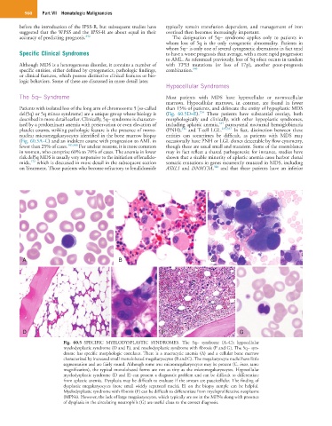
Fig. 60.5 SPECIFIC MYELODYSPLASTIC SYNDROMES. The 5q− syndrome (A–C); hypocellular
myelodysplastic syndrome (D and E), and myelodysplastic syndrome with fibrosis (F and G). The 5q− syn-
drome has specific morphologic correlates. There is a macrocytic anemia (A) and a cellular bone marrow
characterized by increased small monolobated megakaryocytes (B and C). The megakaryocyte nuclei have little
segmentation and are fairly round. Although some true micromegakaryocytes may be present (C, inset, same
magnification), the typical monolobated forms are not as tiny as the micromegakaryocytes. Hypocellular
myelodysplastic syndrome (D and E) can present a diagnostic problem and can be difficult to differentiate
from aplastic anemia. Dysplasia may be difficult to evaluate if the smears are paucicellular. The finding of
dysplastic megakaryocytes (note small widely separated nuclei, E) on the biopsy sample can be helpful.
Myelodysplastic syndrome with fibrosis (F) can be difficult to differentiate from myeloproliferative neoplasms
(MPNs). However, the lack of large megakaryocytes, which typically are see in the MPNs along with presence
of dysplasia in the circulating neutrophils (G) are useful clues to the correct diagnosis.