
Page 1311 - Hematology_ Basic Principles and Practice ( PDFDrive )
P. 1311
Chapter 71 Eosinophilia, Eosinophil-Associated Diseases, Eosinophilic Leukemias, and the Hypereosinophilic Syndromes 1157
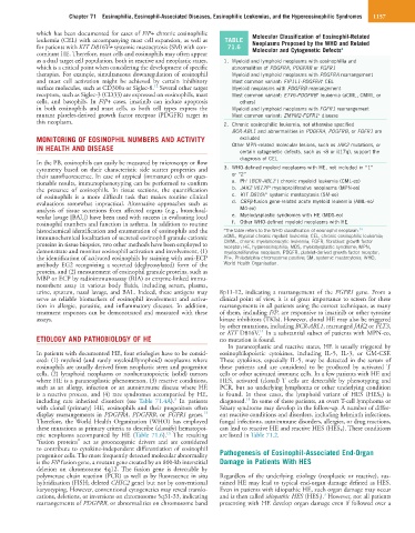
which has been documented for cases of F/P+ chronic eosinophilic Molecular Classification of Eosinophil-Related
leukemia (CEL) with accompanying mast cell expansion, as well as TABLE Neoplasms Proposed by the WHO and Related
for patients with KIT D816V+ systemic mastocytosis (SM) with con- 71.6 Molecular and Cytogenetic Defects a
comitant HE. Therefore, mast cells and eosinophils may often appear
as a dual target cell population, both in reactive and neoplastic states, 1. Myeloid and lymphoid neoplasms with eosinophilia and
which is a critical point when considering the development of specific abnormalities of PDGFRA, PDGFRB or FGFR1
therapies. For example, simultaneous downregulation of eosinophil Myeloid and lymphoid neoplasms with PDGFRA rearrangement
and mast cell activation might be achieved by certain inhibitory Most common variant: FIP1L1-PDGFRA CEL
+
12
surface molecules, such as CD300a or Siglec-8. Several other target Myeloid neoplasms with PDGFRB rearrangement
receptors, such as Siglec-3 (CD33) are expressed on eosinophils, mast Most common variant: ETV6-PDGFRB leukemia (aCML, CMML or
+
cells, and basophils. In F/P+ cases, imatinib can induce apoptosis others)
in both eosinophils and mast cells, as both cell types express the Myeloid and lymphoid neoplasms with FGFR1 rearrangement
mutant platelet-derived growth factor receptor (PDGFR) target in Most common variant: ZMYM2-FGFR1 disease
+
this neoplasm. 2. Chronic eosinophilic leukemia, not otherwise specified
BCR-ABL1 and abnormalities in PDGFRA, PDGFRB, or FGFR1 are
MONITORING OF EOSINOPHIL NUMBERS AND ACTIVITY excluded
IN HEALTH AND DISEASE Other MPN-related molecular lesions, such as JAK2 mutations, or
certain cytogenetic defects, such as +8 or i(17q), support the
diagnosis of CEL
In the PB, eosinophils can easily be measured by microscopy or flow
cytometry based on their characteristic side scatter properties and 3. WHO-defined myeloid neoplasms with HE, not included in “1”
their autofluorescence. In case of atypical (immature) cells or ques- or “2” + +
tionable results, immunophenotyping can be performed to confirm a. Ph (BCR-ABL1 ) chronic myeloid leukemia (CML-eo)
+
the presence of eosinophils. In tissue sections, the quantification b. JAK2 V617F myeloproliferative neoplasms (MPN-eo)
+
of eosinophils is a more difficult task that makes routine clinical c. KIT D816V systemic mastocytosis (SM-eo)
evaluations somewhat impractical. Alternative approaches such as d. CBFβ-fusion gene-related acute myeloid leukemia (AML-eo/
analysis of tissue secretions from affected organs (e.g., bronchoal- M4-eo)
veolar lavage [BAL]) have been used with success in evaluating local e. Myelodysplastic syndromes with HE (MDS-eo)
eosinophil numbers and function in asthma. In addition to routine f. Other WHO-defined myeloid neoplasms with HE
histochemical identification and enumeration of eosinophils and the a The table refers to the WHO classification of eosinophil neoplasm. 14
immunochemical localization of secreted eosinophil granule cationic aCML, Atypical chronic myeloid leukemia; CEL, chronic eosinophilic leukemia;
proteins in tissue biopsies, two other methods have been employed to CMML, chronic myelomonocytic leukemia; FGFR, fibroblast growth factor
receptor; HE, hypereosinophilia; MDS, myelodysplastic syndrome; MPN,
demonstrate and monitor eosinophil activation and involvement, (1) myeloproliferative neoplasm; PDGFR, platelet-derived growth factor receptor;
the identification of activated eosinophils by staining with anti-ECP Ph+, Philadelphia chromosome positive; SM, systemic mastocytosis; WHO,
antibody EG2 recognizing a secreted (deglycosylated) form of the World Health Organisation.
protein, and (2) measurement of eosinophil granule proteins, such as
MBP or ECP by radioimmunoassay (RIA) or enzyme-linked immu-
nosorbent assay in various body fluids, including serum, plasma,
urine, sputum, nasal lavage, and BAL. Indeed, these antigens may 8p11-12, indicating a rearrangement of the FGFR1 gene. From a
serve as reliable biomarkers of eosinophil involvement and activa- clinical point of view, it is of great importance to screen for these
tion in allergic, parasitic, and inflammatory diseases. In addition, rearrangements in all patients using the correct techniques, as many
treatment responses can be demonstrated and measured with these of them, including F/P, are responsive to imatinib or other tyrosine
assays. kinase inhibitors (TKIs). However, clonal HE may also be triggered
by other mutations, including BCR-ABL1, rearranged JAK2 or FLT3,
14
or KIT D816V. In a substantial subset of patients with MPN-eo,
ETIOLOGY AND PATHOBIOLOGY OF HE no mutation is found.
In paraneoplastic and reactive states, HE is usually triggered by
In patients with documented HE, four etiologies have to be consid- eosinophilopoietic cytokines, including IL-5, IL-3, or GM-CSF.
ered: (1) myeloid (and rarely myeloid/lymphoid) neoplasms where These cytokines, especially IL-5, may be detected in the serum of
eosinophils are usually derived from neoplastic stem and progenitor these patients and are considered to be produced by activated T
cells, (2) lymphoid neoplasms or nonhematopoietic (solid) tumors cells or other activated immune cells. In a few patients with HE and
where HE is a paraneoplastic phenomenon, (3) reactive conditions, HES, activated (clonal) T cells are detectable by phenotyping and
such as an allergy, infection or an autoimmune disease where HE PCR, but no underlying lymphoma or other underlying condition
is a reactive process, and (4) rare syndromes accompanied by HE, is found. In these cases, the lymphoid variant of HES (HES L ) is
3
15
including rare inherited disorders (see Table 71.4A). In patients diagnosed. In some of these patients, an overt T-cell lymphoma or
with clonal (primary) HE, eosinophils and their progenitors often Sézary syndrome may develop in the follow-up. A number of differ-
13
display rearrangements in PDGFRA, PDGFRB, or FGFR1 genes. ent reactive conditions and disorders, including helminth infections,
Therefore, the World Health Organization (WHO) has employed fungal infections, autoimmune disorders, allergies, or drug reactions,
these mutations as primary criteria to describe (classify) hematopoi- can lead to reactive HE and reactive HES (HES R ). These conditions
14
etic neoplasms accompanied by HE (Table 71.6). The resulting are listed in Table 71.2.
“fusion proteins” act as prooncogenic drivers and are considered
to contribute to cytokine-independent differentiation of eosinophil
progenitor cells. The most frequently detected molecular abnormality Pathogenesis of Eosinophil-Associated End-Organ
is the F/P fusion gene, a mutant gene created by an 800-kb interstitial Damage in Patients With HES
deletion on chromosome 4q12. The fusion gene is detectable by
polymerase chain reaction (PCR) as well as by fluorescence in situ Regardless of the underlying etiology (neoplastic or reactive), sus-
hybridization (FISH; deleted CHIC2 gene) but not by conventional tained HE may lead to typical end-organ damage defined as HES.
karyotyping. However, conventional cytogenetics may reveal translo- Even in patients with idiopathic HE, such organ damage may occur
3
cations, deletions, or inversions on chromosome 5q31-33, indicating and is then called idiopathic HES (HES I ). However, not all patients
rearrangements of PDGFRB, or abnormalities on chromosome band presenting with HE develop organ damage even if followed over a