
Page 1404 - Hematology_ Basic Principles and Practice ( PDFDrive )
P. 1404
1250 Part VII Hematologic Malignancies
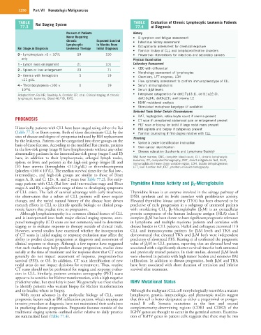
TABLE Rai Staging System TABLE Evaluation of Chronic Lymphocytic Leukemia Patients
77.3 77.4 at Diagnosis
Percent of Patients History
Never Requiring • B symptom and fatigue assessment
Chronic Expected Survival • Infectious history assessment
Lymphocytic in Months From • Occupational assessment for chemical exposure
Rai Stage at Diagnosis Leukemia Therapy Initial Diagnosis
• Familial history of CLL and lymphoproliferative disorders
9
0 - Lymphocytosis >5 × 10 /L 59 150 • Preventive interventions for infections and secondary cancers
only Physical Examination
1 - Lymph node enlargement 21 101 Laboratory Assessment
• CBC with differential
2 - Spleen or liver enlargement 23 71
• Morphology assessment of lymphocytes
3 - Anemia with hemoglobin 5 19 • Chemistry, LFT enzymes, LDH
<11 g/dL • Flow cytometry assessment to confirm immunophenotype of CLL
4 - Thrombocytopenia <100 × 0 19 • Serum immunoglobulins
12
10 /L • Serum β 2 M levels
Adapted from Rai KR, Sawitsky A, Cronkite EP, et al. Clinical staging of chronic • Interphase cytogenetics for del(17p13.1), del(11q22.3),
lymphocytic leukemia, Blood 46:219, 1975. del(13q14), del(6q21), and trisomy 12
• IGHV mutational analysis
• Stimulated metaphase karyotype (if available)
Selected Tests Under Certain Circumstances
• DAT, haptoglobin, reticulocyte count if anemia present
PROGNOSIS • CT scan if unexplained abdominal pain or enlargement present
• PET scan or biopsy (or both) if large nodal mass present
Historically, patients with CLL have been staged using either the Rai • BM aspirate and biopsy if cytopenias present
(Table 77.3) or Binet system. Both of these discriminate CLL by the • Familial counseling if first-degree relative with CLL
sites of disease and degree of cytopenias induced by BM replacement Teaching
by the leukemia. Patients can be categorized into three groups on the • Varicella zoster identification instruction
basis of these features. According to the modified Rai criteria, patients • Skin cancer identification
in the low-risk group (stage 0) have lymphocytosis without any other • Disease education (Leukemia and Lymphoma Society)
abnormality; patients in the intermediate-risk group (stages I and II)
have, in addition to their lymphocytosis, enlarged lymph nodes, BM, Bone marrow; CBC, complete blood count; CLL, chronic lymphocytic
spleen, or liver; and patients in the high-risk group (stages III and leukemia; CT, computed tomography; DAT, direct antiglobulin test; IGHV,
immunoglobulin heavy chain variable region; LDH, lactate dehydrogenase;
IV) have anemia (hemoglobin <11.0 g/dL) or thrombocytopenia LFT, liver function test; PET, positron emission tomography.
9
(platelets <100 × 10 /L). The median survival times for the Rai low-,
intermediate-, and high-risk groups are similar to those of Binet
stages A, B, and C: 12+, 8, and 2 years (see Table 77.2). For early-
stage patients with CLL (Rai low- and intermediate-stage and Binet Thymidine Kinase Activity and β 2-Microglobulin
stages A and B), a significant range of time to developing symptoms
of CLL exists. The lack of survival advantage with early treatment, Thymidine kinase is an enzyme involved in the salvage pathway of
the observation that a subset of CLL patients will never require DNA synthesis and its levels correlate with proliferative activity.
therapy, and the varied natural history of the disease have driven Elevated thymidine kinase activity (TKA) has been observed to be
research efforts in CLL to identify specific biologic or clinical prog- predictive of early progression in a subgroup of untreated patients
nostic factors that predict time to progression. with smoldering CLL. β 2 -Microglobulin (β 2 M) is an extracellular
Although lymphadenopathy is a common clinical feature of CLL protein component of the human leukocyte antigen (HLA) class I
and is incorporated into both major clinical staging systems, com- complex. β 2 M has been shown to have significant prognostic relevance
puted tomography (CT) scans are not commonly used to determine in lymphoma and multiple myeloma patients and correlates with
staging or to evaluate response to therapy outside of clinical trials. disease burden in CLL patients. Hallek and colleagues examined 113
However, several studies have examined whether the incorporation CLL and immunocytoma patients for β 2 M levels and TKA and
of CT scans in initial staging or response evaluation may affect the demonstrated that elevated TKA and β 2 M both were independent
ability to predict disease progression at diagnosis and assessment of predictors of shortened PFS. Keating et al confirmed the prognostic
clinical response to therapy. Although a few reports have suggested value of β 2 M in CLL patients, reporting that an elevated level was
that such studies may help predict disease progression, studies done associated with a significantly shorter survival time for both untreated
serially at the time of treatment and for response show that CT scans and previously treated patients. In their studies, elevated β 2 M levels
generally do not impact assessment of response, progression-free were observed in patients with high tumor burden and extensive BM
survival (PFS), or OS. In addition, CT scan identification of new infiltration. In addition to disease progression, both β 2 M and TKA
nodal areas do not impact decisions for retreatment. Thus, routine have been associated with short duration of remission and inferior
CT scans should not be performed for staging and response evalua- survival after treatment.
tion in CLL. Similarly, positron emission tomography (PET) scans
appear to be sensitive for Richter transformation, with a high negative
predictive value, but specificity is poor. We generally use these studies IGHV Mutational Status
to identify patients who warrant biopsy for Richter transformation
and to localize where to biopsy. Although the malignant CLL cell morphologically resembles a mature
With recent advances in the molecular biology of CLL, some lymphocyte, genetic, immunologic, and phenotypic studies suggest
prognostic factors such as BM infiltration pattern, which requires an that this cell is better designated as either a pregerminal or postger-
invasive procedure at diagnosis, have not maintained their usefulness minal B cell. Somatic mutations in the first and second
in predicting disease progression. Prognostic features outside of the complementarity-determining regions (CDR1 and CDR2) of the
traditional staging systems outlined earlier relative to daily practice IGHV genes are thought to occur in the germinal centers. Examina-
are summarized later (Table 77.4). tion of IGHV genes in patient cells suggests that there may be two