
Page 1433 - Hematology_ Basic Principles and Practice ( PDFDrive )
P. 1433
Chapter 79 Marginal Zone Lymphomas (Extranodal/Malt, Splenic, and Nodal) 1279
autoimmune disorders 60–64 (Table 79.2), although the strength of this similar to those of centrocytes and having relatively abundant pale
correlation for some primary sites of disease is discordant among cytoplasm (leading possibly to a monocytoid appearance). These cells
studies, suggesting a possible geographic variation. The common are located in the outer zone of reactive lymphoid follicles, extend
assumption about this association is that continual immune stimula- into the interfollicular region, and may sometimes colonize the ger-
tion by bacterial or self-antigens leads to expansion of lymphoid minal centers. Larger, immunoblast- or centroblast-like cells may be
elements in the connective tissue adjacent to the epithelium involved, present in small numbers, but an abundance of these should raise
initially leading to a process of reactive lymphoid hyperplasia. Persis- suspicion for diffuse large B-cell lymphoma (DLBCL), which requires
tent lymphocytic activation and proliferation predisposes to the different management. According to the newest WHO classification,
accumulation of genetic errors that ultimately may result in antigen- the term high-grade MALT lymphoma, denoting the presence of sheets
independent growth and, consequently, lymphoma emergence. 65,66 of transformed cells, should not be used and instead these tumors
The histologic distinction between the reactive inflammatory process should be diagnosed as DLBCL.
associated with chronic infection (or autoimmunity) and lymphoma Often there are lymphoid infiltrates invading and destroying
proper may be difficult, in which case demonstration of immuno- glandular structures, with eosinophilic degeneration of epithelial cells
globulin gene monoclonality by molecular studies may aid in estab- (so-called lymphoepithelioid lesions), which are strongly suggestive,
lishing the diagnosis of lymphoma. 67 albeit not pathognomonic, of progression to lymphoma in cases
where the differential diagnosis with reactive lymphoid hyperplasia is
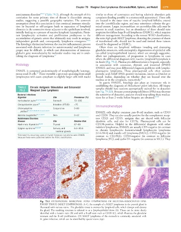
in doubt (Fig. 79.2). Plasmacytic differentiation is frequent, especially
Histology in association with cutaneous, thyroid, and intestinal (IPSID)
ENMZL and may pose differential diagnosis problems with lympho-
ENMZL is composed predominantly of morphologically heteroge- plasmacytic lymphoma. These plasmacytoid cells often contain
67
neous small B cells. These resemble a spectrum spanning from small periodic acid–Schiff (PAS)–positive inclusions, known as Dutcher or
lymphocytes with scant cytoplasm to slightly larger cells with nuclei Russell bodies, depending on whether they are located over the
nucleus or in the cytoplasm, respectively. 68
In gastric ENMZL, histology also plays an important role in
establishing the diagnosis of Helicobacter pylori infection. All biopsy
TABLE Chronic Antigenic Stimulation and Extranodal samples should have sections appropriately stained for its detection
79.2 Marginal Zone Lymphoma (see Fig. 79.2D). Because proton pump inhibitors (PPIs) may decrease
Bacterial Infections the sensitivity of detection, patients should stop taking these medica-
Organism Site Prevalence (%) tions for at least 2 weeks before biopsies are obtained. 69,70
Helicobacter pylori 15,17,41,42,43 Stomach 72–100
Campylobacter jejuni 44 Intestine (IPSID) ≈70 Immunophenotype
Chlamydophila Conjunctiva 0–89
psittaci 45,46,47,48,49,50,51,52,53,54,55 ENMZL cells display common pan–B-cell markers, such as CD19
Borrelia burgdorferi 56,57,58,59 Skin 0–42 and CD20. They are also usually positive for the complement recep-
Autoimmune Disorders tors CD21 and CD35, antigens that are shared with follicular
Disease Site Relative Risk dendritic cells, and also for CD79a. Plasmacytoid cells can be
Hashimoto thyroiditis a61,62 Thyroid 67–80 CD138-positive. Helpful in the differential diagnosis with other
Sjögren syndrome 60,63,64 Salivary and 6.6–30.6 indolent lymphomas, ENMZL are usually CD5-negative (in contrast
lacrimal glands to chronic lymphocytic leukemia/small lymphocytic lymphoma
a Estimated by assuming cases of thyroid histiocytic lymphoma were ENMZL [CLL/SLL] and mantle cell lymphoma [MCL]), CD23-negative (in
contrast to CLL/SLL), CD10-negative (in contrast to follicular
because these were published before the REAL classification.
lymphoma [FL]), and cyclin D1-negative (in contrast to MCL). They
A B C D
Fig. 79.2 EXTRANODAL MARGINAL ZONE LYMPHOMAS OF MUCOSA-ASSOCIATED LYM-
PHOID TISSUE (MALT LYMPHOMAS). A–C, An example of a MALT lymphoma in the parotid gland is
illustrated with various stains. The glandular tissue is overrun by lymphoid cells, which disrupt and destroy
the gland. The resulting structure is referred to as a lymphoepithelial lesion (A). These can be more clearly
identified with a keratin stain (B) and with a B-cell stain such as CD20 (C), which illustrate the glandular
remnant and the B-cell proliferation. (D) MALT lymphoma of the stomach is commonly associated with
H. pylori infection, which can be identified by special stains (top).