
Page 1428 - Hematology_ Basic Principles and Practice ( PDFDrive )
P. 1428
1274 Part VII Hematologic Malignancies
a b
4000 4000
Hairy cell leukemia
Normal B cells
3000 3000 Total cells
SSC-A 2000 SSC-A 2000
1000 1000
0 0
0 10 2 10 3 10 4 10 5 0 10 2 10 3 10 4 10 5
CD20 CD45
c d e
10 5 10 5 10 5
10 4 10 4 10 4
CD11C 10 3 CD25 10 3 CD103 10 3
10 2 10 2 10 2
0 0 0
0 10 2 10 3 10 4 10 5 0 10 2 10 3 10 4 10 5 0 10 2 10 3 10 4 10 5
A CD22 CD19 CD19
Hairy cell leukemia
Normal B cells
Other lymphocytes
a b c
10 5 10 5 10 5
10 4 10 4 10 4
CD11C 10 3 CD25 10 3 CD103 10 3
10 2 10 2 10 2
0 0 0
0 10 2 10 3 10 4 10 5 0 10 2 10 3 10 4 10 5 0 10 2 10 3 10 4 10 5
B CD22 CD19 CD19
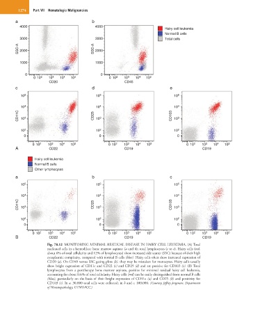
Fig. 78.12 MONITORING MINIMAL RESIDUAL DISEASE IN HAIRY CELL LEUKEMIA. (A) Total
nucleated cells in a hemodilute bone marrow aspirate (a and b); total lymphocytes (c to e). Hairy cells (red,
about 6% of total cellularity and 12% of lymphocytes) show increased side scatter (SSC) because of their high
cytoplasmic complexity, compared with normal B cells (blue). Hairy cells often show increased expression of
CD20 (a). On CD45 versus SSC gating plots (b), they may be mistaken for monocytes. Hairy cells usually
show bright expression of CD11c and CD22 (c) and CD25 (d) and are positive for CD103 (e). (B) Total
lymphocytes from a posttherapy bone marrow aspirate, positive for minimal residual hairy cell leukemia,
accounting for about 0.6% of total cellularity. Hairy cells (red) can be easily distinguished from normal B cells
(blue), particularly on the basis of their bright expression of CD11c (a) and CD25 (b) and positivity for
CD103 (c). In a, 30,000 total cells were collected; in b and c, 100,000. (Courtesy Jeffrey Jorgensen, Department
of Hematopathology, UTMDACC.)