
Page 1558 - Hematology_ Basic Principles and Practice ( PDFDrive )
P. 1558
Chapter 86 Plasma Cell Neoplasms 1385
plasma cells, and these changes have been correlated with an effect
on overall clinical outcome.
MicroRNAs (miRNAs or miRs) are a class of small noncoding
1 2 3 4 5 RNAs that cleave specific targeted transcripts inhibiting translational
of specific genes. Differences in the expression patterns of miRNAs
have been observed between MM and MGUS. For example, miR-32,
and miR-17-92, are overexpressed only in MM, whereas miR-21,
6 7 8 9 10 11 12 miR-106b-25, and miR-181a/b show similar expression patterns in
both MM and MGUS but are highly expressed compared with
normal plasma cells, providing a possible clue to events underlying
13 14 15 16 17 18 progression from MGUS to MM. miRs 15a/16 are present on
chromosome 13, and their downregulation is described in a subset
of MM. Although this does not strictly correlate with chromosome
13 deletion, which is observed in almost half of the patients with
19 20 21 22 X Y MM, a potential effect of these miRs on MM cell proliferation has
been described. A similar attempt at correlating observed cytogenetic
12 changes and their effects on miR expression has identified overexpres-
sion of miR-let-7e, miR-125-5p, and miR-99b in patients with
t(4;14) translocation and miR-1 and miR-133a in patients with
1 2 3 4 5 t(14;16) MM. A causal relationship between changes in these miR
17 1 expression patterns and their effects on target genes and eventual
8
phenotypic changes in MM still needs to be established. As in gene
7
expression profiling, miRNA expression profiling also identifies
6 7 8 9 10 11 12 subgroups with different clinical outcomes, highlighting a significant
role of miRNAs in determining MM biology and its possible role as
a therapeutic target. An integrated analysis of mRNA and miR profil-
13 14 15 16 17 18 ing has been used to identify regulatory networks that combine
1 1 miRNA-mRNA pairs that may drive the behavior of tumor cells. One
1 such network combines p53-MDM2 expression with the downregu-
19 20 21 22 X Y lation of miR-192, miR-194, and miR-215 in a subset of patients
with MM. Such analyses might explain some of the observed genomic
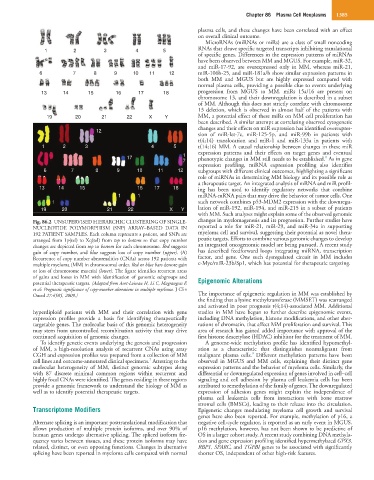
Fig. 86.2 UNSUPERVISED HIERARCHIC CLUSTERING OF SINGLE- changes in myelomagenesis and its progression. Further studies have
NUCLEOTIDE POLYMORPHISM (SNP) ARRAY–BASED DATA IN reported a role for miR-21, miR-29, and miR-34a in supporting
192 PATIENT SAMPLES. Each column represents a patient, and SNPs are myeloma cell and survival, suggesting their potential as novel thera-
arranged from 1p(tel) to Xq(tel) from top to bottom so that copy number peutic targets. Efforts to combine various genomic changes to develop
changes are depicted from top to bottom for each chromosome. Red suggests an integrated oncogenomic model are being pursued. A recent study
gain of copy number, and blue suggests loss of copy number (upper). (A) has described feedforward loops integrating miRNA, transcription
Recurrence of copy number abnormalities (CNAs) across 192 patients with factor, and gene. One such dysregulated circuit in MM includes
multiple myeloma (MM) in chromosomal order. Red or blue bars denote gain c-Myc/miR-23b/Sp1, which has potential for therapeutic targeting.
or loss of chromosome material (lower). The figure identifies recurrent areas
of gains and losses in MM with identification of genomic subgroups and
potential therapeutic targets. (Adapted from Avet-Loiseau H, Li C, Magrangeas F, Epigenomic Alterations
et al: Prognostic significance of copy-number alterations in multiple myeloma. J Clin
Oncol 27:4585, 2009.) The importance of epigenetic regulation in MM was established by
the finding that a lysine methyltransferase (MMSET) was rearranged
and activated in poor prognosis t(4;14)-associated MM. Additional
hyperdiploid patients with MM and their correlation with gene studies in MM have begun to further describe epigenomic events,
expression profiles provide a basis for identifying therapeutically including DNA methylation, histone modifications, and other aber-
targetable genes. The molecular basis of this genomic heterogeneity rations of chromatin, that affect MM proliferation and survival. This
may stem from uncontrolled recombination activity that may drive area of research has gained added importance with approval of the
continued acquisition of genomic changes. first histone deacetylase (HDAC) inhibitor for the treatment of MM.
To identify genetic events underlying the genesis and progression A genome-wide methylation profile has identified hypomethyl-
of MM, a high-resolution analysis of recurrent CNAs using array ation as a characteristic that distinguishes nonmalignant from
9
CGH and expression profiles was prepared from a collection of MM malignant plasma cells. Different methylation patterns have been
7
cell lines and outcome-annotated clinical specimens. Attesting to the observed in MGUS and MM cells, explaining their distinct gene
molecular heterogeneity of MM, distinct genomic subtypes along expression patterns and the behavior of myeloma cells. Similarly, the
with 87 discrete minimal common regions within recurrent and differential or downregulated expression of genes involved in cell–cell
highly focal CNAs were identified. The genes residing in these regions signaling and cell adhesion by plasma cell leukemia cells has been
provide a genomic framework to understand the biology of MM as attributed to remethylation of the family of genes. The downregulated
well as to identify potential therapeutic targets. expression of adhesion genes might explain the independence of
plasma cell leukemia cells from interactions with bone marrow
stromal cells (BMSCs), leading to their release into the circulation.
Transcriptome Modifiers Epigenetic changes modulating myeloma cell growth and survival
genes have also been reported. For example, methylation of p16, a
Alternate splicing is an important posttranslational modification that negative cell-cycle regulator, is reported as an early event in MGUS.
allows production of multiple protein isoforms, and over 90% of p16 methylation, however, has not been shown to be predictive of
human genes undergo alternative splicing. The spliced isoform fre- OS in a larger cohort study. A recent study combining DNA methyla-
quency varies between tissues, and these protein isoforms may have tion and gene expression profiling identified hypermethylated GPX3,
related, distinct, or even opposing functions. Changes in alternative RBP1, SPARC, and TGFBI genes to be associated with significantly
splicing have been reported in myeloma cells compared with normal shorter OS, independent of other high-risk features.