
Page 1596 - Hematology_ Basic Principles and Practice ( PDFDrive )
P. 1596
1422 Part VII Hematologic Malignancies
suggesting that WM cells require a microenvironmental support Unlike most indolent lymphomas, splenomegaly and lymphadenopa-
60
system for their growth and survival. High levels of CXCR4 and thy are uncommon (≤15%). Purpura is frequently associated with
59
very late antigen-4 (VLA-4) are expressed by WM cells. In blocking cryoglobulinemia and in rare circumstances with light-chain (AL)
experiment studies, CXCR4 was shown to support migration of WM amyloidosis. Hemorrhagic and neuropathic manifestations are
cells, whereas VLA-4 contributed to adhesion of WM cells to BM multifactorial (see “IgM-Related Neuropathy” section below). The
stromal cells. 59 morbidity associated with WM is caused by the co-occurrence of two
main components: tissue infiltration by neoplastic cells and, impor-
tantly, the physicochemical and immunologic properties of the
CLINICAL FEATURES monoclonal IgM. As shown in Table 87.2, the monoclonal IgM can
produce clinical manifestations through several different mechanisms
Table 87.1 provides the clinical and laboratory features at the time related to its physicochemical properties, nonspecific interactions
16
of diagnosis of patients with WM in one large institutional study. with other proteins, antibody activity, and tendency to deposit in
tissues. 61–63
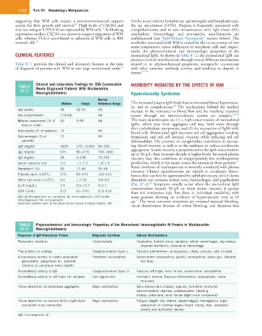
Clinical and Laboratory Findings for 356 Consecutive MORBIDITY MEDIATED BY THE EFFECTS OF IGM
TABLE Newly Diagnosed Patients With Waldenström
87.1
Macroglobulinemia Hyperviscosity Syndrome
Normal
Median Range Reference Range The increased plasma IgM levels lead to increased blood hyperviscos-
64
ity and its complications. The mechanisms behind the marked
Age (years) 58 32–91 NA increase in the resistance to blood flow and the resulting impaired
Sex (male/female) 215/141 NA transit through the microcirculatory system are complex. 64–67
Marrow involvement (% of 30 5–95 NA The main determinants are (1) a high concentration of monoclonal
area on slide) IgMs, which may form aggregates and may bind water through
their carbohydrate component; and (2) the interaction of IgMs with
Adenopathy (% of patients) 15 NA
blood cells. Monoclonal IgM increases red cell aggregation (rouleau
Splenomegaly (% of 10 NA formation) and red cell internal viscosity while reducing red cell
patients) deformability. The presence of cryoglobulins contributes to increas-
IgM (mg/dL) 2620 270–12,400 40–230 ing blood viscosity, as well as to the tendency to induce erythrocyte
aggregation. Serum viscosity is proportional to the IgM concentration
IgG (mg/dL) 674 80–2770 700–1600
up to 30 g/L, then increases sharply at higher levels. Increased plasma
IgA (mg/dL) 58 6–438 70–400 viscosity may also contribute to inappropriately low erythropoietin
67
Serum viscosity (cp) 2.0 1.1–7.2 1.4–1.9 production, which is the major reason for anemia in these patients.
Hematocrit (%) 35 17–45 35–44 Renal synthesis of erythropoietin is inversely correlated with plasma
viscosity. Clinical manifestations are related to circulatory distur-
9
Platelet count (×10 /L) 275 42–675 155–410 bances that can best be appreciated by ophthalmoscopy, which shows
9
White cell count (×10 /L) 6.4 1.7–22 3.8–9.2 distended and tortuous retinal veins, hemorrhages, and papilledema
68
β 2 -M (mg/dL) 2.5 0.9–13.7 0–2.7 (Fig. 87.4). Symptoms usually occur when the monoclonal IgM
concentration exceeds 50 g/L or when serum viscosity is greater
LDH (U/mL) 313 61–1701 313–618 than 4.0 centipoises (cp), but there is individual variability, with
β 2 M, β 2 -Microglobulin; cp, centipoise; Ig, immunoglobulin; LDH, lactate some patients showing no evidence of hyperviscosity even at 10
dehydrogenase; NA, not applicable. cp. The most common symptoms are oronasal mucosal bleeding,
64
Data from patients seen at the Dana-Farber Cancer Institute, Boston, MA.
visual disturbances because of retinal bleeding, and dizziness that
TABLE Physicochemical and Immunologic Properties of the Monoclonal Immunoglobulin M Protein in Waldenström
87.2 Macroglobulinemia
Properties of IgM Monoclonal Protein Diagnostic Condition Clinical Manifestations
Pentameric structure Hyperviscosity Headaches, blurred vision, epistaxis, retinal hemorrhages, leg cramps,
impaired mentation, intracranial hemorrhage
Precipitation on cooling Cryoglobulinemia (type I) Raynaud phenomenon, acrocyanosis, ulcers, purpura, cold urticaria
Autoantibody activity to myelin-associated Peripheral neuropathies Sensorimotor neuropathies, painful neuropathies, ataxic gait, bilateral
glycoprotein, ganglioside M 1 , sulfatide foot drop
moieties on peripheral nerve sheaths
Autoantibody activity to IgG Cryoglobulinemia (type II) Purpura, arthralgia, renal failure, sensorimotor neuropathies
Autoantibody activity to red blood cell antigens Cold agglutinins Hemolytic anemia, Raynaud phenomenon, acrocyanosis, livedo
reticularis
Tissue deposition as amorphous aggregates Organ dysfunction Skin: bullous skin disease, papules, Schnitzler syndrome
Gastrointestinal: diarrhea, malabsorption, bleeding
Kidney: proteinuria, renal failure (light-chain component)
Tissue deposition as amyloid fibrils (light-chain Organ dysfunction Fatigue, weight loss, edema, hepatomegaly, macroglossia, organ
component most commonly) dysfunction of involved organs (heart, kidney, liver, peripheral
sensory and autonomic nerves)
IgM, Immunoglobulin M.