
Page 1862 - Hematology_ Basic Principles and Practice ( PDFDrive )
P. 1862
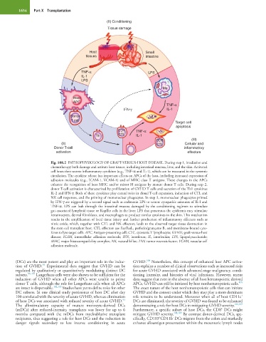
1654   Part X  Transplantation


                                                  (II) Conditioning
                                                   Tissue damage


                                          Host                      Small
                                        tissues                     intestine


                                      TNF-α                          LPS
                                      IL-1
                                      LPS
                                                                            Mφ

                                   Host
                                   APC
                                                                              TNF-α
                                                          IFN-γ               IL-1
                                 Donor
                                  T cell
                                                                                  Target cell
                                                                                  apoptosis
                                                       CD4      TNF-α
                                             Th        CTL      IL-1
                                                                         CD8             (III)
                             (II)                                        CTL          Cellular and
                          Donor T-cell                                                inflammatory
                           activation                                                  effectors

                        Fig. 108.2  PATHOPHYSIOLOGY OF GRAFT-VERSUS-HOST DISEASE. During step 1, irradiation and
                        chemotherapy both damage and activate host tissues, including intestinal mucosa, liver, and the skin. Activated
                        cell hosts then secrete inflammatory cytokines (e.g., TNF-α and IL-1), which can be measured in the systemic
                        circulation. The cytokine release has important effects on APCs of the host, including increased expression of
                        adhesion molecules (e.g., ICAM-1, VCAM-1) and of MHC class II antigens. These changes in the APCs
                        enhance the recognition of host MHC and/or minor H antigens by mature donor T cells. During step 2,
                        donor T-cell activation is characterized by proliferation of GVHD T cells and secretion of the Th1 cytokines
                        IL-2 and IFN-γ. Both of these cytokines play central roles in clonal T-cell expansion, induction of CTL and
                        NK cell responses, and the priming of mononuclear phagocytes. In step 3, mononuclear phagocytes primed
                        by IFN-γ are triggered by a second signal such as endotoxin LPS to secrete cytopathic amounts of IL-I and
                        TNF-α.  LPS  can  leak  through  the  intestinal  mucosa  damaged  by  the  conditioning  regimen  to  stimulate
                        gut-associated lymphoid tissue or Kupffer cells in the liver; LPS that penetrates the epidermis may stimulate
                        keratinocytes, dermal fibroblasts, and macrophages to produce similar cytokines in the skin. This mechanism
                        results in the amplification of local tissue injury and further production of inflammatory effectors such as
                        nitric oxide, which, together with CTL and NK effectors, leads to the observed target tissue destruction in
                        the stem cell transplant host. CTL effectors use Fas/FasL, perforin/granzyme B, and membrane-bound cyto-
                        kines to lyse target cells. APC, Antigen-presenting cell; CTL, cytotoxic T lymphocyte; GVHD, graft-versus-host
                        disease;  ICAM,  intercellular  adhesion  molecule;  IFN,  interferon;  IL,  interleukin;  LPS,  lipopolysaccharide;
                        MHC, major histocompatibility complex; NK, natural killer; TNF, tumor necrosis factor; VCAM, vascular cell
                        adhesion molecule.


                                                                    104
        (DCs) are the most potent and play an important role in the induc-  GVHD.  Nonetheless, this concept of enhanced host APC activa-
                     95
        tion  of  GVHD.   Experimental  data  suggest  that  GVHD  can  be   tion explains a number of clinical observations such as increased risks
        regulated by qualitatively or quantitatively modulating distinct DC   for acute GVHD associated with advanced-stage malignancy, condi-
        subsets. 96–101  Langerhans cells were also shown to be sufficient for the   tioning intensity, and histories of viral infections. However, recent
        induction  of  GVHD  when  all  other  APCs  were  unable  to  prime   data suggest that even in the absence of all host hematopoietic derived
                                                                                                               105
        donor T cells, although the role for Langerhans cells when all APCs   APCs, GVHD can still be initiated by host nonhematopoietic cells.
        are intact is dispensable. 102,103  Studies have yet to define roles for other   The exact nature of the host nonhematopoietic cells that can initiate
        DC subsets. In one clinical study persistence of host DC after day   GVHD and the context under which they may play a more dominant
                                                                                                                +
        100 correlated with the severity of acute GVHD, whereas elimination   role remains to be understood. Moreover when all of host CD11c
                                                         93
        of host DCs was associated with reduced severity of acute GVHD.    DCs are eliminated, the severity of GVHD was found to be enhanced
        The  allostimulatory  capacity  of  mature  monocyte-derived  DCs   demonstrating a role for host DCs in mitigating GVHD severity. 106,107
                                                                                                       +
        (mDCs)  after  reduced-intensity  transplants  was  lower  for  up  to  6   Furthermore, a specific subset of host DCs, the CD8  DCs might
        months  compared  with  the  mDCs  from  myeloablative  transplant   mitigate GVHD severity. 108,109  By contrast donor-derived DCs, spe-
                                                                          +
                                                                                -
        recipients, thus suggesting a role for host DCs and the reduction in   cifically, CD103 CD11b  DCs migrate from the colon and markedly
        danger  signals  secondary  to  less  intense  conditioning  in  acute   enhance alloantigen presentation within the mesenteric lymph nodes