
Page 1953 - Hematology_ Basic Principles and Practice ( PDFDrive )
P. 1953
Chapter 113 Human Leukocyte Antigen and Human Neutrophil Antigen Systems 1733
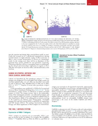
A B
tPE
tPE
CD8FITC CD8FITC
Fig. 113.6 SCHEMATIC REPRESENTATION OF THE MECHANISM OF BINDING OF TETRA-
MERIC PEPTIDE/HUMAN LEUKOCYTE ANTIGEN COMPLEXES (THLA) TO ANTIGEN-SPECIFIC
T CELLS. (A) Binding of tHLA to T-cell receptor. The tHLA consists of four HLA/peptide complexes identical
to the ones recognized by the T cell on the surface of live cells. Each HLA molecule is modified to contain
one biotin molecule that serves as a bridge for binding to tetravalent streptavidin molecules fluorescently
+
labeled. (B) Actual fluorescence-activated cell sorter analysis result of CD8 , tHLA-positive T cells. (Modified
from Monsurro V, Nargosen D: Immunotracking of specific cancer vaccine CD8 lymphocytes. ASHI Q 26:100, 2003.)
into the rejection mechanism when matched-donor grafts are used. TABLE International Society of Blood Transfusion
Finally, the increased use of T cell-directed immunization is driving 113.4 Nomenclature
renewed interest in high-resolution typing of HLA molecules to
allow a more accurate interpretation of clinical and immunologic Antigen Former
results. In the coming years, there will be an abundance of data System Antigens Location Name Alleles
generated from various methods for next generation sequencing. HNA-1 HNA-1a FcγRIIIb, CD16 NA1 FCGR3B*01
Utilization of this data will have a tremendous impact on understand- HNA-1b FcγRIIIb, CD16 NA2 FCGR3B*02
ing the clinical and immunologic implications in regards to HLA. HNA-1c FcγRIIIb, CD16 SH FCGR3B*03
This data will require an enormous effort with bioinformatics at the HNA-1d FcγRIIIb, CD16 FCGR3B*02
forefront.
HNA-2 HNA-2 CD177 (NB1 gp) NB1 CD177
HNA-3 HNA-3a CTL-2 5b SLC44A2*01
HUMAN NEUTROPHIL ANTIGENS AND HNA-4 HNA-4a CD11b (CR3) Mart(a) ITGAM*01
THEIR CLINICAL SIGNIFICANCE HNA-5 HNA-5a CD11a (LFA-1) Ond(a) ITGAL*01
Lalezari et al described the first granulocyte antigens. 214,215 These CR3, C3bi receptor; gp, glycoprotein; HNA, human neutrophil antigen; LFA-1,
leukocyte function–associated antigen-1.
antigens were designated N for neutrophil. Each antigen system was
described alphabetically, and each allele was described numerically
in order of discovery. They identified NA1 in 1966 and its allele,
NA2, in 1972. 214,215 Reports of several other granulocyte antigens
followed. antigens are expressed on all segmented neutrophils, approximately
A new nomenclature was established in 1998 by the International one-half of neutrophilic metamyelocytes, and approximately 10% of
216
221
Society of Blood Transfusion Working Party (Table 113.4). In this neutrophilic myelocytes. Neutrophil expression of FcγRIIIb and
nomenclature, antigen systems are referred to as human neutrophil HNA-1 antigens are diminished by the treatment of neutrophils with
antigens, or HNA. The antigen systems—that is, the polymorphic stimulants such as complement component C5a and chemotaxin
forms of the immunogenic proteins—are indicated by integers, and F-met-leu-phe or granulocyte colony-stimulating factor (G-CSF).
specific antigens within each system are designated alphabetically by Soluble FcγRIIIb is present in plasma, and it has the same HNA-1
date of publication. Alleles of the coding genes are named according polymorphisms found on neutrophils. FcγRIIIb released from
to the Guidelines for Human Gene Nomenclature. Neutrophil granulocytes is the source of the soluble FcγRIIIb and HNA-1
antigens NA1 and NA2 became HNA-1a and HNA-1b, respectively, antigens. 222
and NB1 became HNA-2. The serology, biochemistry, molecular
biology, and clinical significance of these antigens are reviewed here.
Biochemistry
THE HNA-1 ANTIGEN SYSTEM FcγRIIIb is a glycoprotein with 233 amino acids and is glycosylphos-
phatidylinositol (GPI) anchored. 217–220 Its molecular weight is 50 to
Expression of HNA-1 Antigens 80 kDa, and the glycoprotein has N-linked carbohydrate side chains.
The HNA-1a form of FcγRIIIb is 50 to 65 kDa, while the HNA-1b
HNA-1 antigens are expressed only on neutrophils. HNA-1 anti- form of FcγRIIIb is 65 to 80 kDa, and the heterozygous form is 50
gens are located on the low-affinity Fc-γ receptor IIIb (FcγRIIIb), to 80 kDa. Differences in N-glycosylation account for the differences
CD16. 217–220 FcγRIIIb, HNA-1a, HNA-1b, HNA-1c, and HNA-1d in molecular mass.