
Page 204 - Hematology_ Basic Principles and Practice ( PDFDrive )
P. 204
156 Part II Cellular Basis of Hematology
Endothelial
cells Angiogenic gradient formation (“switch”)
Pericytes VEGF Hypoxia, inflammation, oncogenic transformation
Increased expression of stimulators (VEGF)
Basement Decreased expression of inhibitors
membrane
Endothelial (phalanx) cell stimulation
VEGF
Basement membrane dissolution
Pericyte "drop out"
Circumferential extension (mother vessels)
Tip cell
VEGFR2 Formation of endothelial tip cells
VEGF VEGF gradient sensing
Expression of tip cell markers
Metabolic adaptation (glycolysis)
Formation of endothelial sprouts
NOTCH DII4 Directional migration of tip and stalk cells
Blockade of VEGFR2 expression on stalk cells
VEGFR2
by tip cells via the Dll4/Notch pathway
Tip guidance (semaphorins, neuropilins, plexins, Robo4)
Extension of sprouts
Phalanx cells Tie2 Growth and migration of stalk cells and lumen formation
Stalk cells NRP1
Tip cells Myeloid cell–dependent anastomosis
Signalling by Tie2, Ang2, PlGF, PHD2, SDF-1a
Formation of new vascular loops
Pericyte
recruitment Connection and anastomosis of sprouts
Vascular maturation
Pericyte recruitment/vessel maturation (PDGFRb, Ang1)
Restoration of basement membrane (TIMPs, PAI-1)
Endothelial quiescence–phalanx cells (Notch)
Junction formation (VE-cadherin)
Anticoagulant surfaces
Blood flow
New capillary
Resolution of hypoxia
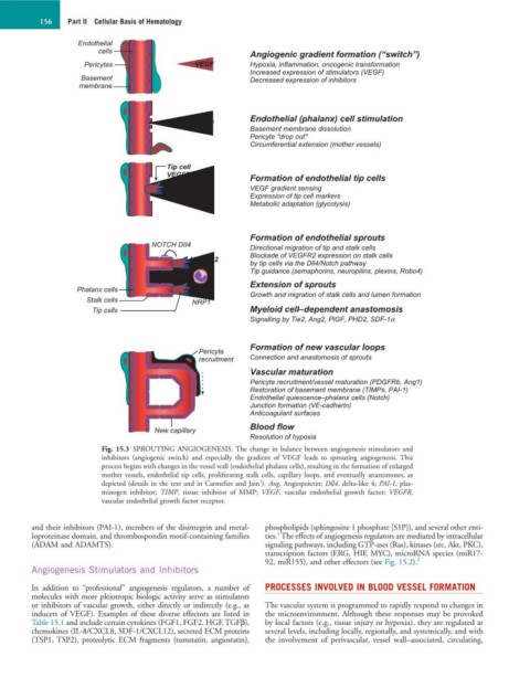
Fig. 15.3 SPROUTING ANGIOGENESIS. The change in balance between angiogenesis stimulators and
inhibitors (angiogenic switch) and especially the gradient of VEGF leads to sprouting angiogenesis. This
process begins with changes in the vessel wall (endothelial phalanx cells), resulting in the formation of enlarged
mother vessels, endothelial tip cells, proliferating stalk cells, capillary loops, and eventually anastomoses, as
2
depicted (details in the text and in Carmeliet and Jain ). Ang, Angiopoietin; Dll4, delta-like 4; PAI-1, plas-
minogen inhibitor; TIMP, tissue inhibitor of MMP; VEGF, vascular endothelial growth factor; VEGFR,
vascular endothelial growth factor receptor.
and their inhibitors (PAI-1), members of the disintegrin and metal- phospholipids (sphingosine 1 phosphate [S1P]), and several other enti-
1
loproteinase domain, and thrombospondin motif-containing families ties. The effects of angiogenesis regulators are mediated by intracellular
(ADAM and ADAMTS). signaling pathways, including GTP-ases (Ras), kinases (src, Akt, PKC),
transcription factors (ERG, HIF, MYC), microRNA species (miR17-
92, miR155), and other effectors (see Fig. 15.2). 2
Angiogenesis Stimulators and Inhibitors
In addition to “professional” angiogenesis regulators, a number of PROCESSES INVOLVED IN BLOOD VESSEL FORMATION
molecules with more pleiotropic biologic activity serve as stimulators
or inhibitors of vascular growth, either directly or indirectly (e.g., as The vascular system is programmed to rapidly respond to changes in
inducers of VEGF). Examples of these diverse effectors are listed in the microenvironment. Although these responses may be provoked
Table 15.1 and include certain cytokines (FGF1, FGF2, HGF, TGFβ), by local factors (e.g., tissue injury or hypoxia), they are regulated at
chemokines (IL-8/CXCL8, SDF-1/CXCL12), secreted ECM proteins several levels, including locally, regionally, and systemically, and with
(TSP1, TSP2), proteolytic ECM fragments (tumstatin, angiostatin), the involvement of perivascular, vessel wall–associated, circulating,