
Page 2072 - Hematology_ Basic Principles and Practice ( PDFDrive )
P. 2072
Chapter 122 Overview of Hemostasis and Thrombosis 1841
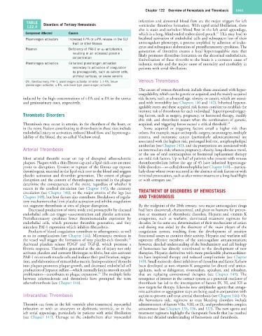
TABLE Disorders of Tertiary Hemostasis infarction and abnormal blood flow are the major triggers for left
122.4 ventricular thrombus formation. With rapid atrial fibrillation, there
also is stasis and turbulent blood flow in the left atrial appendage,
Component Affected Causes which is a long, blind-ended trabeculated pouch. This may lead to
27
Plasminogen activators Increased t-PA or u-PA release in the GU localized activation of endothelial cells and subsequent loss of their
tract or other tissues anticoagulant phenotype, a process amplified by adhesion of leuko-
cytes and subsequent elaboration of proinflammatory cytokines. The
Plasmin Deficiency of PAI-1 or α 2 -antiplasmin, generation of thrombin creates a local hypercoagulable state that
resulting in an increased plasmin likely promotes thrombus formation on the abnormal endothelium.
concentration
Embolization of these thrombi to the brain is a common cause of
Plasminogen activation Enhanced plasminogen activation ischemic stroke and the major cause of mortality and morbidity in
secondary to activation of coagulation patients with atrial fibrillation.
by procoagulants, such as cancer cells,
artificial surfaces, or snake venoms
GU, Genitourinary; PAI-1, plasminogen activator inhibitor 1; t-PA, tissue Venous Thrombosis
plasminogen activator; u-PA, urokinase-type plasminogen activator.
The causes of venous thrombosis include those associated with hyper-
coagulability, which can be genetic or acquired, and the mainly acquired
induced by the high concentrations of t-PA and u-PA in the uterus risk factors, such as advanced age, obesity, or cancer, which are associ-
and genitourinary tract, respectively. ated with immobility (see Chapters 140 and 142). Inherited hyperco-
agulable states and these acquired risk factors combine to establish the
2
intrinsic risk of thrombosis for each individual. Superimposed trigger-
Thrombotic Disorders ing factors, such as surgery, pregnancy, or hormonal therapy, modify
this risk, and thrombosis occurs when the combination of genetic,
Thrombosis may occur in arteries, in the chambers of the heart, or acquired, and triggering forces exceed a critical threshold. 28
in the veins. Factors contributing to thrombosis in these sites include Some acquired or triggering factors entail a higher risk than
endothelial injury or activation, reduced blood flow, and hypercoagu- others. For example, major orthopedic surgery, neurosurgery, multiple
lability of the blood, the so-called Virchow triad. trauma, and metastatic cancer (particularly adenocarcinoma) are
associated with the highest risk; prolonged bed rest, antiphospholipid
antibodies (see Chapter 141), and the puerperium are associated with
Arterial Thrombosis an intermediate risk; whereas pregnancy, obesity, long-distance travel,
or the use of oral contraceptives or hormonal replacement therapy
Most arterial thrombi occur on top of disrupted atherosclerotic are mild risk factors. Up to half of patients who present with venous
plaques. Plaques with a thin fibrous cap and a lipid-rich core are most thromboembolism before the age of 45 have inherited hypercoagu-
prone to disruption. Erosion or rupture of the fibrous cap exposes lable disorders—so-called thrombophilia (see Chapter 140)—particu-
thrombogenic material in the lipid-rich core to the blood and triggers larly those whose event occurred in the absence of risk factors or with
platelet activation and thrombin generation. The extent of plaque minimal provocation, such as after minor trauma or a long-haul flight
disruption and the content of thrombogenic material in the plaque or with estrogen use. 29
determine the consequences of the event, regardless of whether it
occurs in the cerebral circulation (see Chapter 145), the coronary
circulation (see Chapter 146), or the major arteries of the legs (see TREATMENT OF DISORDERS OF HEMOSTASIS
Chapter 148), but host factors also contribute. Breakdown of regula- AND THROMBOSIS
tory mechanisms that limit platelet activation and inhibit coagulation
can augment thrombosis at sites of plaque disruption. By the midpoint of the 20th century, two major anticoagulant drugs
Decreased production of nitric oxide and prostacyclin by diseased had been discovered, characterized, and given to humans for preven-
endothelial cells can trigger vasoconstriction and platelet activation. tion or treatment of thrombotic disorders. Heparin and vitamin K
Proinflammatory cytokines lower thrombomodulin expression by antagonists, such as warfarin, dominated treatment regimens for
endothelial cells, which promotes thrombin generation, and they decades. In the same era, determination of their mechanisms of action
stimulate PAI-1 expression which inhibits fibrinolysis. and dosing was aided by the discovery of the main players of the
Products of blood coagulation contribute to atherogenesis, as well coagulation system, resulting from the development of sensitive
as to its complications (see Chapter 144). Microscopic erosions in functional assays to monitor their activity. Heparin and warfarin still
26
the vessel wall trigger the formation of tiny platelet-rich thrombi. represent effective members of the anticoagulant armamentarium;
Activated platelets release PDGF and TGF-β, which promote a however, detailed understanding of the biochemistry and cell biology
fibrotic response. Thrombin generated at the site of injury not only of hemostasis has directly contributed to the development of new
activates platelets and converts fibrinogen to fibrin, but also activates therapies. Heparin derivatives with more predictable pharmacokinet-
PAR-1 on smooth muscle cells and induces their proliferation, migra- ics have improved therapy and reduced complications (see Chapter
tion, and elaboration of extracellular matrix. Incorporation of thrombi 149). Small molecule, direct inhibitors of thrombin and factor Xa have
into plaques promotes plaque growth, and decreased endothelial cell been developed as non–vitamin K antagonist (or direct) oral antico-
production of heparan sulfate—which normally limits smooth muscle agulants, such as dabigatran, rivaroxaban, apixaban, and edoxaban,
26
proliferation—contributes to plaque expansion. The multiple links that are replacing conventional therapies (see Chapter 149). The
between atherosclerosis and thrombosis have prompted the term resurgence of interest in the contact system as a potential mediator of
atherothrombosis (see Chapter 144). thrombosis has led to the investigation of factors IX, XI, and XII as
new targets for therapy. Likewise new antiplatelet agents that antago-
nize activation or aggregation steps are being used in conjunction with
Intracardiac Thrombosis aspirin to prevent and treat arterial thrombosis (see Chapter 146). On
the hemostasis side, regimens to treat bleeding disorders include
Thrombi can form in the left ventricle after transmural myocardial administration of factors VIIa, VIII, or IX, and prothrombin complex
infarction or with an aneurysm or dyskinetic ventricle, or in the concentrates (see Chapters 135, 136, and 137). The new agents and
left atrial appendage, particularly in patients with atrial fibrillation treatment regimens highlight the therapeutic benefit that has resulted
(see Chapter 147). Damage to the endothelium after myocardial from our detailed understanding of hemostasis and thrombosis.