
Page 2070 - Hematology_ Basic Principles and Practice ( PDFDrive )
P. 2070
Chapter 122 Overview of Hemostasis and Thrombosis 1839
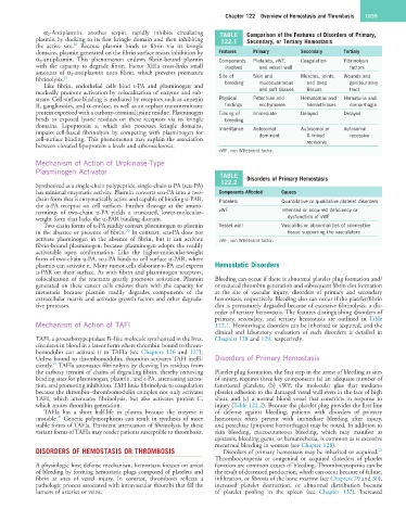
α 2-Antiplasmin, another serpin, rapidly inhibits circulating TABLE Comparison of the Features of Disorders of Primary,
plasmin by docking to its first kringle domain and then inhibiting 122.1 Secondary, or Tertiary Hemostasis
20
the active site. Because plasmin binds to fibrin via its kringle
domains, plasmin generated on the fibrin surface resists inhibition by Features Primary Secondary Tertiary
α 2-antiplasmin. This phenomenon endows fibrin-bound plasmin Components Platelets, vWF, Coagulation Fibrinolysis
with the capacity to degrade fibrin. Factor XIIIa cross-links small involved and vessel wall factors
amounts of α 2-antiplasmin onto fibrin, which prevents premature
fibrinolysis. 19 Site of Skin and Muscles, joints, Wounds and
Like fibrin, endothelial cells bind t-PA and plasminogen and bleeding mucocutaneous and deep genitourinary
markedly promote activation by colocalization of enzyme and sub- and soft tissues tissues tract
strate. Cell-surface binding is mediated by receptors such as annexin Physical Petechiae and Hematomas and Hematuria and
II, gangliosides, and α-enolase, as well as an orphan transmembrane findings ecchymoses hemarthroses menorrhagia
protein expressed with a carboxy-terminal lysine residue. Plasminogen Timing of Immediate Delayed Delayed
binds to exposed lysine residues on these receptors via its kringle bleeding
domains. Lipoprotein a, which also possesses kringle domains, Inheritance Autosomal Autosomal or Autosomal
impairs cell-based fibrinolysis by competing with plasminogen for dominant X-linked recessive
cell-surface binding. This phenomenon may explain the association recessive
between elevated lipoprotein a levels and atherosclerosis.
vWF, von Willebrand factor.
Mechanism of Action of Urokinase-Type
Plasminogen Activator
TABLE Disorders of Primary Hemostasis
122.2
Synthesized as a single-chain polypeptide, single-chain u-PA (scu-PA)
has minimal enzymatic activity. Plasmin converts scu-PA into a two- Components Affected Causes
chain form that is enzymatically active and capable of binding u-PAR, Platelets Quantitative or qualitative platelet disorders
the u-PA receptor on cell surfaces. Further cleavage at the amino-
terminus of two-chain u-PA yields a truncated, lower-molecular- vWF Inherited or acquired deficiency or
weight form that lacks the u-PAR binding domain. dysfunction of vWF
Two-chain forms of u-PA readily convert plasminogen to plasmin Vessel wall Vasculitis or abnormalities of connective
20
in the absence or presence of fibrin. In contrast, scu-PA does not tissue supporting the vasculature
activate plasminogen in the absence of fibrin, but it can activate vWF, von Willebrand factor.
fibrin-bound plasminogen, because plasminogen adopts the readily
activatable open conformation. Like the higher-molecular-weight
form of two-chain u-PA, scu-PA binds to cell surface u-PAR, where
plasmin can activate it. Many tumor cells elaborate u-PA and express Hemostatic Disorders
u-PAR on their surface. As with fibrin and plasminogen receptors,
colocalization of the reactants greatly promotes activation. Plasmin Bleeding can occur if there is abnormal platelet plug formation and/
generated on these cancer cells endows them with the capacity for or reduced thrombin generation and subsequent fibrin clot formation
metastasis because plasmin readily degrades components of the at the site of vascular injury; disorders of primary and secondary
extracellular matrix and activates growth factors and other degrada- hemostasis, respectively. Bleeding also can occur if the platelet/fibrin
tive proteases. clot is prematurely degraded because of excessive fibrinolysis; a dis-
order of tertiary hemostasis. The features distinguishing disorders of
primary, secondary, and tertiary hemostasis are outlined in Table
Mechanism of Action of TAFI 122.1. Hemorrhagic disorders can be inherited or acquired, and the
clinical and laboratory evaluation of such disorders is detailed in
TAFI, a procarboxypeptidase B–like molecule synthesized in the liver, Chapters 128 and 129, respectively.
circulates in blood in a latent form where thrombin bound to throm-
bomodulin can activate it to TAFIa (see Chapters 126 and 127).
Unless bound to thrombomodulin, thrombin activates TAFI ineffi- Disorders of Primary Hemostasis
21
ciently. TAFIa attenuates fibrinolysis by cleaving Lys residues from
the carboxy termini of chains of degrading fibrin, thereby removing Platelet plug formation, the first step in the arrest of bleeding at sites
binding sites for plasminogen, plasmin, and t-PA, attenuating activa- of injury, requires three key components (a) an adequate number of
tion, and promoting inhibition. TAFI links fibrinolysis to coagulation functional platelets, (b) vWF, the molecular glue that mediates
because the thrombin–thrombomodulin complex not only activates platelet adhesion to the damaged vessel wall even in the face of high
TAFI, which attenuates fibrinolysis, but also activates protein C, shear, and (c) a normal blood vessel that constricts in response to
which mutes thrombin generation. injury (Table 122.2). Because the platelet plug provides the first line
TAFIa has a short half-life in plasma because the enzyme is of defense against bleeding, patients with disorders of primary
21
unstable. Genetic polymorphisms can result in synthesis of more hemostasis often present with immediate bleeding after injury,
stable forms of TAFIa. Persistent attenuation of fibrinolysis by these and petechiae (pinpoint hemorrhages) may be noted. In addition to
variant forms of TAFIa may render patients susceptible to thrombosis. skin bleeding, mucocutaneous bleeding, which may manifest as
epistaxis, bleeding gums, or hematochezia, is common as is excessive
menstrual bleeding in women (see Chapter 128).
DISORDERS OF HEMOSTASIS OR THROMBOSIS Disorders of primary hemostasis may be inherited or acquired.
22
Thrombocytopenia or congenital or acquired disorders of platelet
A physiologic host defense mechanism, hemostasis focuses on arrest function are common causes of bleeding. Thrombocytopenia can be
of bleeding by forming hemostatic plugs composed of platelets and the result of decreased production, which can occur because of failure,
fibrin at sites of vessel injury. In contrast, thrombosis reflects a infiltration, or fibrosis of the bone marrow (see Chapters 29 and 30),
pathologic process associated with intravascular thrombi that fill the increased platelet destruction, or abnormal distribution because
lumens of arteries or veins. of platelet pooling in the spleen (see Chapter 132). Increased