
Page 2069 - Hematology_ Basic Principles and Practice ( PDFDrive )
P. 2069
1838 Part XII Hemostasis and Thrombosis
COOH NH 2 NH 2 COOH
Aα FPA FPA Aα
Bβ FPB FPB Bβ Fibrinogen
γ γ
D domain Colied E domain Colied D domain
coil coil
D E D Fibrinogen
Thrombin FPA, FPB
D E D Fibrin monomer
D E D D E D
Fibrin polymer
E D D E D D E
Factor XIIIa
D E D D E D Cross-linked
fibrin polymer
E D D E D D E
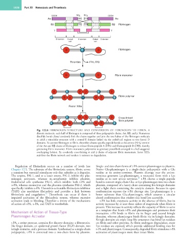
Fig. 122.6 FIBRINOGEN STRUCTURE AND CONVERSION OF FIBRINOGEN TO FIBRIN. A
dimeric molecule, each half of fibrinogen is composed of three polypeptide chains, Aα, Bβ, and γ. Numerous
disulfide bonds (lines) covalently link the chains together and join the two halves of the fibrinogen molecule
to yield a trinodular structure with a central E domain linked via the coiled-coil regions to two lateral D
domains. To convert fibrinogen to fibrin, thrombin cleaves specific peptide bonds at the amino (NH 2) termini
of the Aα and Bβ chains of fibrinogen to release fibrinopeptide A (FPA) and fibrinopeptide B (FPB), thereby
generating fibrin monomer. Fibrin monomers polymerize to generate protofibrils arranged in a half-staggered
overlapping fashion. By covalently cross-linking α and γ chains of adjacent fibrin monomers, factor XIIIa
stabilizes the fibrin network and renders it resistant to degradation.
Regulation of fibrinolysis occurs on a number of levels (see Single- and two-chain forms of t-PA convert plasminogen to plasmin.
Chapter 127). The substrate of the fibrinolytic system, fibrin, serves Native Glu-plasminogen is a single-chain polypeptide with a Glu
a transient but essential stimulatory role that subsides as it degrades. residue at its amino-terminus. Plasmin cleavage near the amino-
The serpins, PAI-1, and to a lesser extent, PAI-2, inhibit the plas- terminus generates Lys-plasminogen, a truncated form with a Lys
20
minogen activators, whereas α 2 -antiplasmin inhibits plasmin. residue at its new amino terminus. t-PA cleaves a single peptide
Endothelial cells synthesize PAI-1, which inhibits both t-PA and bond to convert single-chain Glu- or Lys-plasminogen into two-chain
u-PA, whereas monocytes and the placenta synthesize PAI-2, which plasmin, composed of a heavy chain containing five kringle domains
specifically inhibits u-PA. Thrombin-activatable fibrinolysis inhibitor and a light chain containing the catalytic domain. Because its open
(TAFI) also modulates fibrinolysis and provides a link between conformation exposes the t-PA cleavage site, Lys-plasminogen is a
21
fibrinolysis and coagulation. Thrombosis can occur if there is better substrate than Glu-plasminogen, which assumes a circular
impaired activation of the fibrinolytic system, whereas excessive closed conformation that renders this bond less accessible.
activation leads to bleeding. Therefore a review of the mechanisms t-PA has little enzymatic activity in the absence of fibrin, but its
of action of t-PA, u-PA, and TAFI is worthwhile. activity increases by at least three orders of magnitude when fibrin is
present. This increase in activity reflects the capacity of fibrin to serve
Mechanism of Action of Tissue-Type as a template that binds t-PA and plasminogen and promotes their
interaction. t-PA binds to fibrin via its finger and second kringle
Plasminogen Activator domains, whereas plasminogen binds fibrin via its kringle domains.
Kringle domains are triple loop-like structures that bind Lys residues
t-PA, a serine protease, contains five discrete domains: a fibronectin- on fibrin and other proteins. As fibrin undergoes degradation, more
like finger domain, an epidermal growth factor (EGF) domain, two Lys residues are exposed, which provide additional binding sites for
kringle domains, and a protease domain. Synthesized as a single-chain t-PA and plasminogen. Consequently, degraded fibrin stimulates t-PA
polypeptide, t-PA is converted into a two-chain form by plasmin. activation of plasminogen more than intact fibrin.