
Page 221 - Hematology_ Basic Principles and Practice ( PDFDrive )
P. 221
Chapter 16 Cytokine/Receptor Families and Signal Transduction 173
TABLE Consequence of Deficiencies in Genes of Intracellular
SOCS 16.1 Signaling Molecules
A- SOCS KIR SH2
box
Signal
Transduction
Molecule Phenotype of Deficient Mice
JAK1 Perinatal mortality, defects in IL-6, IL-2, and cytokine
receptor type II families
JAK2 Embryonic lethality caused by defective definitive
hematopoiesis. Defects in TPO, IL-2 family,
Sumo binding
IL-3 and IFN-γ signaling
JAK3 Immunodeficiency because of absent common γ chain
signaling. JAK3 expression is restricted to the
hematopoietic system
B- PIAS SAP SP-RING S/T
Tyk2 Reduced responses to IFN-α/β, IL-12, and unexpectedly
IFN-γ
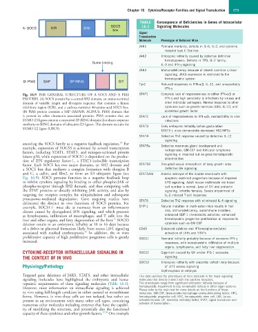
Fig. 16.9 THE GENERAL STRUCTURE OF A SOCS AND A PIAS STAT1 Complete lack of responsiveness to either IFN-α/β or
PROTEIN. (A) SOCS protein has a central SH2 domain, an amino-terminal IFN-γ and high sensitivity to infections by viruses and
domain of variable length and divergent sequence that contains a kinase other microbial pathogens. Normal response to other
inhibitory region (KIR), and a carboxy-terminal 40-amino-acid SOCS box. cytokines such as growth hormone (GH), IL-10, and
(B) PIAS protein contains a SAP (SAFA/B, ACINUS, PIAS) domain that epidermal growth factor
is present in other chromatin associated proteins. PIAS proteins that are STAT2 Lack of responsiveness to IFN-α/β; susceptibility to viral
SUMO E3 ligases contain a conserved SP-RING domain that shares sequence infections
similarity to RING domains of ubiquitin E3 ligases. This domain recruits the STAT3 Early embryonic lethality before gastrulation
SUMO E2 ligase (UBC9).
STAT3 ± mice demonstrate decreased HSC/HPCs
STAT4 Defective Th1 response caused by defective IL-12
signaling
34
encoding the SOCS family as a negative feedback regulation. For STAT5a Defective mammary gland development and
example, expression of SOCS3 is activated by several transcription lactogenesis; GM-CSF and follicular lymphoma
factors, including STAT1, STAT3, and mitogen-activated protein signaling is impaired but no gross hematopoietic
kinase p38, while expression of SOCS1 is dependent on the produc- abnormalities
tion of IFN regulatory factor-1, a STAT1-inducible transcription
factor. Each SOCS has two major domains, an SH2 domain and STAT5B Disrupted sexual dimorphism of body growth rates.
a SOCS box that mediates a complex formation with elongins B Defective GH signaling
and C, a cullin, and Rbx2, to form an E3 ubiquitin ligase (see STAT5A/B Anemic embryos of the double knock-outs with
Fig. 16.9). SOCS proteins function in a negative feedback loop apoptotic erythroid progenitors because of impaired
to inhibit cytokine signaling by binding to either phospho-JAK or EPO signaling. Adult mouse erythrocyte red blood
phospho-receptor through SH2 domain, and thus competing with cell number is normal. Loss of GH and prolactin
the STAT proteins or directly inhibiting JAK activity, and also by signaling. Infertile females. Severe impairment of
targeting the receptor complex for ubiquitylation and subsequent IL-2 induced T-cell responses
proteasome-mediated degradation. Gene targeting studies have STAT6 Defective Th2 response with eliminated IL-4 signaling
delineated the distinct in vivo functions of SOCS proteins. For
example, SOCS1-/- mice die as neonates from an inflammatory SHP-1 Natural mutation in moth-eaten mice results in hair
disease caused by dysregulated IFN signaling, and which presents loss, immunodeficiency, autoimmune disorders,
as lymphopenia, infiltration of macrophages, and T cells into the enhanced SDF-1 chemotactic activities, enhanced
35
liver and other organs, and fatty degeneration of the liver. SOCS3 hematopoietic progenitor proliferation in response to
deletion results in an embryonic lethality at 10 to 16 days because cytokines such as GM-CSF
of a defect in placental formation likely from excess LIF1 signaling CD45 Enhanced cytokine and IFN-receptor-mediated
36
associated with marked erythrocytosis. In addition, the in vitro activation of JAKs and STATs
proliferative capacity of high proliferative progenitor cells is greatly SOCS1 Neonatal lethality probably because of excessive IFN-γ
increased. responses, with hematopoietic infiltration of multiple
organs, lymphopenia, and fatty liver degeneration
CYTOKINE-RECEPTOR INTRACELLULAR SIGNALING IN SOCS2 Gigantism caused by GH and/or IFG-1 excessive
THE CONTEXT OF IN VIVO signaling
SOCS3 Embryonic lethality with placental defect likely because
Physiology/Pathology of LIF1 excess signaling
Erythrocytosis in embryos
Targeted gene deletions of JAKS, STATS, and other intracellular This table outlines the phenotypes of mice deficient in the major signaling
signaling molecules have highlighted the embryonic and hema- molecules that directly interact with the cytokine receptors.
topoietic requirements of these signaling molecules (Table 16.1). The phenotypes range from significant embryonic lethality because of
However, most information on intracellular signaling is achieved hematopoietic impairment to less remarkable defects in other organ systems.
Please refer to the main text for more details on the functions of these
ex vivo using full-length cytokines in either natural or recombinant molecules. GM-CSF, Granulocyte-macrophage colony-stimulating factor; HPC,
forms. However, in vivo these cells are not isolated, but rather are hematopoietic progenitor cell; HSC, hematopoietic stem cell; JAK, Janus-
present in an environment with many other cell types, containing activated kinase; LIF, leukemia inhibitory factor; STAT, signal transducer and
numerous other molecules including enzymes that have the capabil- activator of transcription.
ity of modifying the structure, and potentially also the functional
capacity of these cytokines and other growth factors. 37,38 One example