
Page 2347 - Hematology_ Basic Principles and Practice ( PDFDrive )
P. 2347
Chapter 141 The Antiphospholipid Syndrome 2089
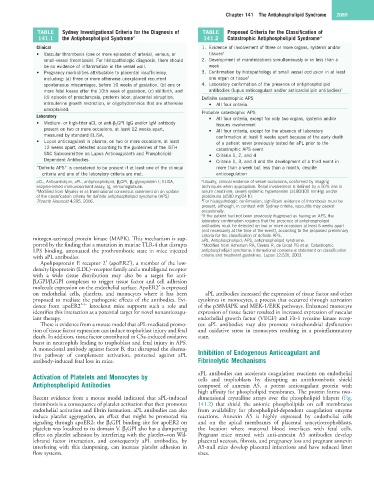
TABLE Sydney Investigational Criteria for the Diagnosis of TABLE Proposed Criteria for the Classification of
141.1 the Antiphospholipid Syndrome a 141.2 Catastrophic Antiphospholipid Syndrome*
Clinical 1. Evidence of involvement of three or more organs, systems and/or
• Vascular thrombosis (one or more episodes of arterial, venous, or tissues a
small-vessel thrombosis). For histopathologic diagnosis, there should 2. Development of manifestations simultaneously or in less than a
be no evidence of inflammation in the vessel wall. week
• Pregnancy morbidities attributable to placental insufficiency, 3. Confirmation by histopathology of small vessel occlusion in at least
including: (a) three or more otherwise unexplained recurrent one organ or tissue b
spontaneous miscarriages, before 10 weeks of gestation, (b) one or 4. Laboratory confirmation of the presence of antiphospholipid
more fetal losses after the 10th week of gestation, (c) stillbirth, and antibodies (lupus anticoagulant and/or anticardiolipin antibodies) c
(d) episode of preeclampsia, preterm labor, placental abruption, Definite catastrophic APS
intrauterine growth restriction, or oligohydramnios that are otherwise • All four criteria
unexplained. Probable catastrophic APS
Laboratory • All four criteria, except for only two organs, systems and/or
• Medium- or high-titer aCL or anti-β 2 GPI IgG and/or IgM antibody tissues involvement
present on two or more occasions, at least 12 weeks apart, • All four criteria, except for the absence of laboratory
measured by standard ELISA. confirmation at least 6 weeks apart because of the early death
• Lupus anticoagulant in plasma, on two or more occasions, at least of a patient never previously tested for aPL prior to the
12 weeks apart, detected according to the guidelines of the ISTH catastrophic APS event
SSC Subcommittee on Lupus Anticoagulants and Phospholipid- • Criteria 1, 2, and 4
Dependent Antibodies. • Criteria 1, 3, and 4 and the development of a third event in
“Definite APS” is considered to be present if at least one of the clinical more than a week but less than a month, despite
criteria and one of the laboratory criteria are met. anticoagulation
aCL, Anticardiolipin; aPL, antiphospholipid; β 2 GPI, β 2 -glycoprotein I; ELISA, a Usually, clinical evidence of vessel occlusions, confirmed by imaging
enzyme-linked immunosorbent assay; Ig, immunoglobulin. techniques when appropriate. Renal involvement is defined by a 50% rise in
a Modified from Miyakis et al: International consensus statement on an update serum creatinine, severe systemic hypertension (≥180/100 mmHg) and/or
of the classification criteria for definite antiphospholipid syndrome (APS). proteinuria (≥500 mg/24 h).
Thromb Haemost 4:295, 2006. b For histopathologic confirmation, significant evidence of thrombosis must be
present, although, in contrast with Sydney criteria, vasculitis may coexist
occasionally.
c If the patient had not been previously diagnosed as having an APS, the
laboratory confirmation requires that the presence of antiphospholipid
antibodies must be detected on two or more occasions at least 6 weeks apart
(not necessarily at the time of the event), according to the proposed preliminary
criteria for the classification of definite APS.
mitogen-activated protein kinase (MAPK). This mechanism is sup- aPL, Antiphospholipid; APS, antiphospholipid syndrome.
ported by the finding that a mutation in murine TLR-4 that disrupts *Modified from Asherson RA, Cevera R, de Groot PG et al. Catastrophic
LPS binding, attenuated the prothrombotic state in mice injected antiphospholipid syndrome: international consensus statement on classification
with aPL antibodies. criteria and treatment guidelines. Lupus 12:530, 2003.
Apolipoprotein E receptor 2′ (apoER2′), a member of the low-
density lipoprotein (LDL)–receptor family and a multiligand receptor
with a wide tissue distribution may also be a target for anti-
β 2 GPI/β 2 GPI complexes to trigger tissue factor and cell adhesion
molecule expression on the endothelial surface. ApoER2′ is expressed
on endothelial cells, platelets, and monocytes where it has been aPL antibodies increased the expression of tissue factor and other
proposed to mediate the pathogenic effects of the antibodies. Evi- cytokines in monocytes, a process that occurred through activation
–/–
dence from apoER2′ knockout mice supports such a role and of the p38MAPK and MEK-1/ERK pathways. Enhanced monocyte
identifies this interaction as a potential target for novel nonanticoagu- expression of tissue factor resulted in increased expression of vascular
lant therapy. endothelial growth factor (VEGF) and Flt-1 tyrosine kinase recep-
There is evidence from a mouse model that aPL-mediated promo- tor. aPL antibodies may also promote mitochondrial dysfunction
tion of tissue factor expression can induce trophoblast injury and fetal and oxidative stress in monocytes resulting in a proinflammatory
death. In addition, tissue factor contributed to C5a-induced oxidative state.
burst in neutrophils leading to trophoblast and fetal injury in APS.
A monoclonal antibody against factor B, that disrupted the alterna-
tive pathway of complement activation, protected against aPL Inhibition of Endogenous Anticoagulant and
antibody-induced fetal loss in mice. Fibrinolytic Mechanisms
Activation of Platelets and Monocytes by aPL antibodies can accelerate coagulation reactions on endothelial
cells and trophoblasts by disrupting an antithrombotic shield
Antiphospholipid Antibodies composed of annexin A5, a potent anticoagulant protein with
high affinity for phospholipid membranes. The protein forms two-
Recent evidence from a mouse model indicated that aPL-induced dimensional crystalline arrays over the phospholipid bilayers (Fig.
thrombosis is a consequence of platelet activation that then promotes 141.2) that shield the anionic phospholipids on cell membranes
endothelial activation and fibrin formation. aPL antibodies can also from availability for phospholipid-dependent coagulation enzyme
induce platelet aggregation, an effect that might be promoted via reactions. Annexin A5 is highly expressed by endothelial cells
signaling through apoER2; the β 2GPI binding site for apoER2 on and on the apical membranes of placental syncytiotrophoblasts,
platelets was localized to its domain V. β 2 GPI also has a dampening the location where maternal blood interfaces with fetal cells.
effect on platelet adhesion by interfering with the platelet–von Wil- Pregnant mice treated with anti-annexin A5 antibodies develop
lebrand factor interaction, and consequently aPL antibodies, by placental necrosis, fibrosis, and pregnancy loss and pregnant annexin
interfering with this dampening, can increase platelet adhesion in A5-null mice develop placental infarctions and have reduced litter
flow systems. sizes.