
Page 2349 - Hematology_ Basic Principles and Practice ( PDFDrive )
P. 2349
Chapter 141 The Antiphospholipid Syndrome 2091
TABLE Proposed Pathogenic Mechanisms of Antiphospholipid Syndrome
141.3
I. aPL-mediated promotion of tissue factor expression
A. Direct injury and subsequent anti-β 2 GPI binding on endothelial cells
B. Signaling via annexin A2/TLR4/apoER2 inducing proadhesive prothrombotic phenotype
C. Induction of adhesion molecules and tissue factor on endothelial cells and cytokine release
II. Activation of platelets and monocytes by aPL antibodies
A. Activation of platelets: via apoER2′, GPIbα, and/or β 2 GPI-platelet factor 4 interaction
B. Interference of β 2 GPI in regulating vWF-mediated platelet adhesion
C. Activation of monocytes: results in increased tissue factor, VEGF, cytokine expression
D. Activation of monocytes causes mitochondrial dysfunction and oxidative stress
III. Inhibition of endogenous anticoagulant and fibrinolytic mechanisms
A. Disruption of the annexin A5 anticoagulant shield
B. Interference with fibrinolysis via annexin A2, β 2 GPI cofactor activity, autoactivation of XIIa, direct inhibition of plasmin and increase of PAI-1
C. Inhibition of the protein C pathway: decreased activation of protein C, barrier of APC proteolysis of factor Va and VIIIa, prevention of protein C
and EPCR binding
D. Interference with tissue factor pathway inhibitor
IV. aPL-mediated activation of complement
A. Antibodies against β 2 GPI–HLA-DR7 complexes on cell surfaces trigger complement-mediated cytotoxicity
V. Direct activation of trophoblasts and endometrial cells by aPL antibodies
A. Abnormal trophoblast proliferation, migration and invasiveness, increased trophoblast apoptosis, and reduced secretion of HCG and adhesion
molecules
B. Disruption in the differentiation of decidual endometrial cells
C. Disruption of maternal spiral artery transformation and maturation
VI. Other mechanisms
A. mTORC pathway–mediated vasculopathy
B. Release in procoagulant microparticles by endothelial cells and platelets
APC, activated protein C; aPL, antiphospholipid; apoER2; apolipoprotein E receptor 2; EPCR, endothelial cell protein C receptor; β 2 GPI, β 2 -glycoprotein I; HCG, human
chorionic gonadotropin; HLA, human leukocyte antigen; mTORC, mammalian target of rapamycin complex; PAI-1, plasminogen activator inhibitor 1; TLR, Toll-like
receptor; VEGF, vascular endothelial growth factor; vWF, von Willebrand factor
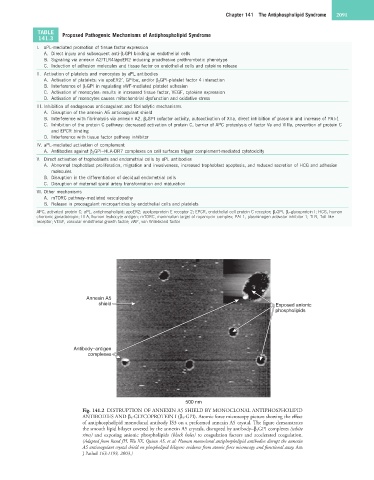
Annexin A5
shield Exposed anionic
phospholipids
Antibody–antigen
complexes
500 nm
Fig. 141.2 DISTRUPTION OF ANNEXIN A5 SHIELD BY MONOCLONAL ANTIPHOSPHOLIPID
ANTIBODIES AND β 2 -GLYCOPROTEIN I (β 2 -GPI). Atomic force microscopy picture showing the effect
of antiphospholipid monoclonal antibody IS3 on a preformed annexin A5 crystal. The figure demonstrates
the smooth lipid bilayer covered by the annexin A5 crystals, disrupted by antibody–β 2 GPI complexes (white
rims) and exposing anionic phospholipids (black holes) to coagulation factors and accelerated coagulation.
(Adapted from Rand JH, Wu XX, Quinn AS, et al: Human monoclonal antiphospholipid antibodies disrupt the annexin
A5 anticoagulant crystal shield on phospholipid bilayers: evidence from atomic force microscopy and functional assay. Am
J Pathol 163:1193, 2003.)