
Page 2421 - Hematology_ Basic Principles and Practice ( PDFDrive )
P. 2421
Chapter 148 Peripheral Artery Disease 2161
stenosis, and an extensive collateral circulation. In such cases, mea-
surement of the ABI after walking will detect a decrease in the ankle
systolic pressure relative to brachial artery systolic pressure, thereby
revealing the presence of PAD.
Noninvasive Testing and Imaging for
Diagnosis of Peripheral Artery Disease
Several other noninvasive tests may help in the diagnosis of PAD and
in the identification of sites of stenosis. These tests include segmental
pressures with pulse volume recordings, duplex ultrasonography,
computed tomography angiography (CTA), magnetic resonance
angiography (MRA), and conventional contrast angiography. When
measuring segmental leg pressures, systolic blood pressure measure-
ments are obtained at multiple levels in the leg, typically in the upper
thigh, lower thigh, upper calf, ankle, and across the metatarsal region
of the foot. Systolic blood pressures in these sites are then compared
with the higher of the arm systolic blood pressures. A significant drop
in blood pressure (>20 mmHg) from one level to the next can localize
arterial stenosis with a high degree of precision. An upper thigh
pressure that is lower than the arm pressure indicates stenosis in the
distal aorta or in the iliac or femoral arteries (or both). In patients
with vascular calcification, measurement and interpretation of seg-
mental pressures are unreliable. Pulse volume recordings can also be
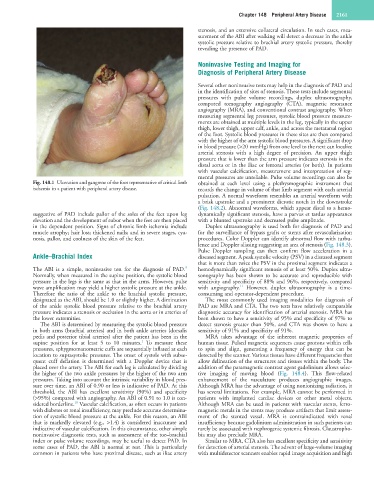
Fig. 148.1 Ulceration and gangrene of the foot representative of critical limb obtained at each level using a plethysmographic instrument that
ischemia in a patient with peripheral artery disease. records the change in volume of that limb segment with each arterial
pulsation. A normal waveform resembles an arterial waveform with
a brisk upstroke and a prominent dicrotic notch in the downstroke
(Fig. 148.2). Abnormal waveforms, which appear distal to a hemo-
suggestive of PAD include pallor of the soles of the feet upon leg dynamically significant stenosis, have a parvus et tardus appearance
elevation and the development of rubor when the feet are then placed with a blunted upstroke and decreased pulse amplitude.
in the dependent position. Signs of chronic limb ischemia include Duplex ultrasonography is used both for diagnosis of PAD and
muscle atrophy; hair loss; thickened nails; and in severe stages, cya- for the surveillance of bypass grafts or stents after revascularization
nosis, pallor, and coolness of the skin of the feet. procedures. Color Doppler can identify abnormal flow with turbu-
lence and Doppler aliasing suggesting an area of stenosis (Fig. 148.3).
Pulse Doppler sampling can then confirm flow acceleration in a
Ankle–Brachial Index diseased segment. A peak systolic velocity (PSV) in a diseased segment
that is more than twice the PSV in the proximal segment indicates a
9
The ABI is a simple, noninvasive test for the diagnosis of PAD. hemodynamically significant stenosis of at least 50%. Duplex ultra-
Normally, when measured in the supine position, the systolic blood sonography has been shown to be accurate and reproducible with
pressure in the legs is the same as that in the arms. However, pulse sensitivity and specificity of 88% and 96%, respectively, compared
11
wave amplification may yield a higher systolic pressure at the ankle. with angiography. However, duplex ultrasonography is a time-
Therefore the ratio of the ankle to the brachial systolic pressure, consuming and operator-dependent procedure.
designated as the ABI, should be 1.0 or slightly higher. A diminution The most commonly used imaging modalities for diagnosis of
of the ankle systolic blood pressure relative to the brachial artery PAD are MRA and CTA. The two tests have relatively comparable
pressure indicates a stenosis or occlusion in the aorta or in arteries of diagnostic accuracy for identification of arterial stenosis. MRA has
the lower extremities. been shown to have a sensitivity of 95% and specificity of 97% to
The ABI is determined by measuring the systolic blood pressure detect stenosis greater than 50%, and CTA was shown to have a
in both arms (brachial arteries) and in both ankle arteries (dorsalis sensitivity of 91% and specificity of 91%.
pedis and posterior tibial arteries) after the patient has been in the MRA takes advantage of the inherent magnetic properties of
1
supine position for at least 5 to 10 minutes. To measure these human tissue. Pulsed magnetic sequences cause protons within cells
pressures, sphygmomanometric cuffs are sequentially inflated at each to spin and align, generating a frequency of energy that can be
location to suprasystolic pressures. The onset of systole with subse- detected by the scanner. Various tissues have different frequencies that
quent cuff deflation is determined with a Doppler device that is allow delineation of the structures and tissues within the body. The
placed over the artery. The ABI for each leg is calculated by dividing addition of the paramagnetic contrast agent gadolinium allows selec-
the higher of the two ankle pressures by the higher of the two arm tive imaging of moving blood (Fig. 148.4). This flow-related
pressures. Taking into account the intrinsic variability in blood pres- enhancement of the vasculature produces angiographic images.
sure over time, an ABI of 0.90 or less is indicative of PAD. At this Although MRA has the advantage of using nonionizing radiation, it
threshold, the ABI has excellent sensitivity (90%) and specificity has several limitations. For example, MRA cannot be performed in
(>95%) compared with angiography. An ABI of 0.91 to 1.0 is con- patients with implanted cardiac devices or other metal objects.
10
sidered borderline. Vascular calcification, as often occurs in patients Although MRA can be used in patients with vascular stents, ferro-
with diabetes or renal insufficiency, may preclude accurate determina- magnetic metals in the stents may produce artifacts that limit assess-
tion of systolic blood pressure at the ankle. For this reason, an ABI ment of the stented vessel. MRA is contraindicated with renal
that is markedly elevated (e.g., >1.4) is considered inaccurate and insufficiency because gadolinium administration in such patients can
indicative of vascular calcification. In this circumstance, other simple rarely be associated with nephrogenic systemic fibrosis. Claustropho-
noninvasive diagnostic tests, such as assessment of the toe–brachial bia may also preclude MRA.
index or pulse volume recordings, may be useful to detect PAD. In Similar to MRA, CTA also has excellent specificity and sensitivity
some cases of PAD, the ABI is normal at rest. This is particularly for detection of arterial stenosis. The advent of large-volume imaging
common in patients who have proximal disease, such as iliac artery with multidetector scanners enables rapid image acquisition and high