
Page 2460 - Hematology_ Basic Principles and Practice ( PDFDrive )
P. 2460
2198 Part XII Hemostasis and Thrombosis
Portal Vein Thrombosis
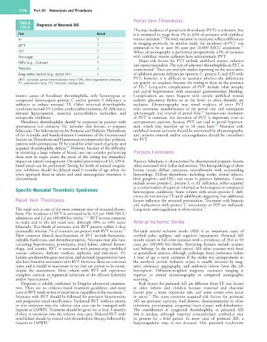
TABLE Diagnosis of Neonatal DIC
150.6
The true incidence of portal vein thrombosis (PVT) is unknown, but
Test Result it is estimated to range from 1% to 43% of neonates with umbilical
30
PT ↑ venous catheters. The wide variation in incidence reflects differences
aPTT ↑ in imaging protocols. In another study, the incidence of PVT was
31
estimated to be at least 36 cases per 10,000 NICU admissions.
TCT ↑ When ultrasonography is performed prospectively, 43% of neonates
Fibrinogen ↓ with umbilical venous catheters have asymptomatic PVT.
Major risk factors for PVT include umbilical venous catheters
FDPs (e.g., D-dimer) ↑
and sepsis/omphalitis. The role of inherited thrombophilia in PVT is
Platelets ↓ controversial. There are multiple studies reporting on the association
Coagulation factors (e.g., factor VIII) ↓ of inhibitor protein deficiencies (protein C, protein S, and AT) with
aPTT, Activated partial thromboplastin time; FDPs, fibrin degradation products; PVT; however, it is difficult to ascertain whether the deficiencies
PT, prothrombin time; TCT, thrombin clotting time. are genetic or acquired, because the testing is done in the presence
30
of TE. Long-term complications of PVT include lobar atrophy
and portal hypertension with associated gastrointestinal bleeding.
known causes of hereditary thrombophilia, only homozygous or Complications are more frequent with ectopic umbilical venous
compound heterozygous protein C and/or protein S deficiency is catheter placement (below or in the liver) or when thrombi are
sufficient to induce neonatal TE. Other inherited thrombophilic occlusive. Ultrasonography may reveal evidence of prior PVT
conditions include FV Leiden, prothrombin mutation, AT deficiency, with cavernous transformation of the portal vein and subsequent
32
elevated lipoprotein(a), maternal anticardiolipin antibodies, and splenomegaly and reversal of portal flow. Spontaneous resolution
nonspecific inhibitors. of PVT is common, but detection of PVT is important, even in
Hereditary thrombophilia should be suspected in patients with asymptomatic patients, because PVT can lead to portal hyperten-
32
spontaneous and extensive TE, ischemic skin lesions, or purpura sion, which may manifest up to 10 years later. Neonates with
fulminans. The Subcommittee for Perinatal and Pediatric Thrombosis umbilical venous catheters should be monitored by ultrasonography,
of the Scientific and Standardization Committee of the International and catheter removal and/or anticoagulation should be considered
Society on Thrombosis and Haemostasis recommended that pediatric for PVT.
patients with spontaneous TE be tested for a full panel of genetic and
28
acquired thrombophilic defects. However, because of the difficulty
in obtaining a large volume of blood, one can consider performing Purpura Fulminans
these tests in stages unless the result of the testing has immediate
impact on patient management. On initial presentation of TE, DNA- Purpura fulminans is characterized by disseminated purpuric lesions
based assays can be performed. Testing for levels of natural coagula- often associated with bullae and necrosis. The histopathology of these
tion inhibitors should be delayed until 6 months of age when the lesions reveals diffuse cutaneous microthrombi with surrounding
levels approach those in adults and until anticoagulant treatment is hemorrhage. Diffuse thrombosis, including stroke, retinal infarcts,
discontinued. limb gangrene, and DIC, can occur in purpura fulminans. Causes
include severe protein C, protein S, or AT deficiency, either acquired
Specific Neonatal Thrombotic Syndromes as a complication of sepsis or inherited as homozygous or compound
heterozygous conditions. Some infants with severe protein C defi-
ciency do not develop TE until adulthood, suggesting that additional
Renal Vein Thrombosis factors influence the neonatal presentation. Treatment with heparin
and replacement with protein C concentrate or FFP are indicated.
The renal vein is one of the most common sites of neonatal throm- Long-term anticoagulation is often needed. 33
bosis. The incidence of RVT is estimated to be 0.5 per 1000 NICU
admissions and 2.2 per 100,000 live births. 23,25 RVT is more common
in males and in the left renal vein, although 28% to 44% occur Arterial Ischemic Stroke
bilaterally. Two-thirds of neonates with RVT present within 3 days
29
postnatally, whereas 7% of neonates can present with RVT in utero. Perinatal arterial ischemic stroke (AIS) is an important cause of
Most common clinical manifestations of RVT include hematuria, cerebral palsy, epilepsy, and cognitive impairment. Perinatal AIS
palpable flank mass, and thrombocytopenia. Neonates may also have mostly occurs in full-term neonates with a prevalence of 28.6 to 93
coexisting hypertension, proteinuria, renal failure, adrenal hemor- cases per 100,000 live births. Presenting features include seizures
rhage, and anemia. RVT is associated with prematurity, umbilical and lethargy. In the neonatal period, AIS often presents with focal
venous catheters, diabetic mothers, asphyxia, and infections. FV or generalized seizures, although pathologic hand preference before
Leiden, prothrombin gene mutation, and elevated lipoprotein(a) have 1 year of age is most common if the stroke was asymptomatic in
also been found in association with RVT. However, these are common the newborn period. Ischemic injury is usually detected by mag-
traits, and it would be inaccurate to say they are proven to be causal, netic resonance angiography, and unilateral lesions favor the left
despite the associations. Most infants with RVT will experience hemisphere. Diffusion-weighted magnetic resonance imaging is
complete, cortical, or segmental infarction of the affected kidney(s) superior to cranial ultrasonography or computed tomographic
and/or hypertension. 21 scanning. 7
Diagnosis is reliably confirmed by Doppler ultrasound examina- Risk factors for perinatal AIS are different from TE risk factors
tion. There are no evidence-based treatment guidelines, and most in older infants and children because maternal and placental
29
cases of RVT result in loss of renal tissue, regardless of the treatment. factors play a more important role, and some events even occur
34
Neonates with RVT should be followed for persistent hypertension in utero. The most common acquired risk factors for perinatal
and progressive renal insufficiency. Unilateral RVT without uremia AIS are perinatal asphyxia, fetal distress, chorioamnionitis or other
or clot extension into the inferior vena cava can be managed with infections, preeclampsia, congenital heart disease, and dehydration.
heparin or LMWH. Treatment should be given for at least 3 months The contribution of congenital thrombophilia to perinatal AIS
if there is extension into the inferior vena cava. Bilateral RVT with risk is unclear, although maternal anticardiolipin antibodies may
renal failure should be treated with thrombolytic therapy followed by be present for a brief period. In most cases of perinatal AIS, a
heparin or LMWH. 7 hypercoagulable state is not detected. One potential mechanism