
Page 2456 - Hematology_ Basic Principles and Practice ( PDFDrive )
P. 2456
2194 Part XII Hemostasis and Thrombosis
Bleeding may be from mucocutaneous sites, sites of capillary blood platelet aggregation in response to all agonists and deficiency of α IIbβ 3
sampling, cephalohematoma, umbilical stump, or at sites of proce- on flow cytometry are diagnostic for Glanzmann thrombasthenia (see
dures. Platelet function disorders result from defects in a number of Chapters 125 and 130). Flow cytometry also provides definitive
structures and signaling pathways as outlined in Table 150.5. Only diagnostic information about Bernard-Soulier syndrome, dense
the most severe genetic disorders of platelet function present in the granule deficiency, and Scott syndrome.
neonatal period. These include Glanzmann thrombasthenia and Platelet transfusions are often given if the patient is bleeding.
Bernard-Soulier syndrome (see Chapters 125 and 130). Maternal However, the potential risk of human leukocyte antigen allosensitiza-
medications may affect platelet function, most notably aspirin, tion if normal platelets are given to patients with congenital deficiency
although low-dose aspirin does not appear to alter neonatal platelet of platelet surface antigens must be weighed against the severity of
function. Neonatal medications may also affect platelet function. bleeding when functional defects are suspected. It is recommended
Common offenders include nitric oxide, prostaglandin E 2 , indo- to restrict platelet transfusion in patients with Glanzmann thrombas-
methacin, and aspirin. thenia. Recombinant activated FVII (rFVIIa) has been used to avoid
It is especially challenging to diagnose platelet function disorders allosensitization. Other adjunctive measures include local control
in neonates because of the technical limitations of many platelet such as use of fibrin sealant in oral bleeding and antifibrinolytic
function assays and the need for large volumes of blood for testing. medications, such as tranexamic acid.
Initial screening for suspected platelet function disorders can be
performed using the PFA-100 analyzer followed by evaluation of
platelet morphology and platelet aggregation responses using light Vitamin K Deficiency Bleeding
transmission aggregometry. Flow cytometry can be used to evaluate
specific surface glycoproteins, and electron microscopy studies can be Vitamin K is an essential cofactor for γ-glutamyl carboxylase, the
used to assess platelet granule morphology. enzyme required for posttranslational carboxylation of prothrombin;
Although the PFA-100 is a sensitive test for detecting hemostatic FVII; FIX; FX; and proteins C, S, and Z. Many newborns are defi-
disorders in the pediatric population, it is relatively nonspecific. Light cient in vitamin K, whether measured in cord blood or indirectly by
transmission aggregometry is highly reproducible in patients with measuring the levels of the vitamin K–dependent coagulation pro-
inherited mucocutaneous bleeding if properly standardized. Absent teins. Risk factors for bleeding with vitamin K deficiency include
maternal malabsorption, maternal intake of drugs that impair vitamin
K metabolism, exclusive breastfeeding, and neonatal malabsorption.
Classification is based on the time of presentation.
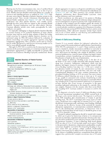
TABLE Inherited Disorders of Platelet Function Early vitamin K deficiency bleeding occurs in the first 24 to
150.5
48 hours of life and is usually associated with maternal intake of
Defects in Receptors for Adhesive Proteins drugs (e.g., phenytoin, barbiturates, antibiotics), which cross the
a
Bernard-Soulier syndrome, platelet-type von Willebrand disease placenta and impair vitamin K synthesis. Classical vitamin K defi-
(GPIb-IX-V complex) ciency bleeding manifests from days 2 to 7 of life and is related to
Glanzmann thrombasthenia (GPIIb/IIIa, α IIb β 3 ) low placental transfer of vitamin K, low concentrations in breast
a
milk, lack of gastrointestinal bacterial flora, and poor oral intake.
GPIa/IIa, α 2 β 1
GPVI Gastrointestinal bleeding is the most common presentation, but
GPIV procedural bleeding, bruising, or ICH can occur. Late-onset vitamin
Defects in Soluble Agonist Receptors K deficiency–related bleeding can occur at times up to 12 weeks
Thromboxane A2 receptor of age, usually in association with exclusive breastfeeding or with
α 2 -Adrenergic receptor neonatal fat malabsorption, and often manifests as catastrophic bleed-
P2Y 12 receptor ing, including ICH. Congenital vitamin K deficiency is an autosomal
Defects in Platelet Granules recessive disorder that occurs because of mutations in the genes
encoding γ-glutamyl carboxylase or the vitamin K 2,3 –epoxide reduc-
δ-Storage pool deficiency, Hermansky-Pudlak syndrome, Chediak- tase complex. Neonates with this disorder often have severe bleed-
Higashi syndrome, thrombocytopenia with absent radii syndrome ing, including ICH. Genotype analysis is necessary to confirm the
(δ-granules) defects. 14
Gray platelet syndrome, Quebec platelet disorder, Paris-Trousseau- The diagnostic criteria for vitamin K deficiency bleeding include
Jacobsen syndrome (α-granules) an elevated international normalized ratio (INR), normal levels of
α,δ-storage pool deficiency (α- and δ-granules) fibrinogen, and a normal platelet count. The diagnosis is confirmed
Defects in Signal-Transduction Pathways if the INR normalizes after administration of vitamin K and the
Primary secretion defects bleeding stops. The aPTT is prolonged with severe vitamin K defi-
Abnormalities of the arachidonic acid/TXA2 pathway ciency. Prophylaxis with a single dose of vitamin K (0.5 to 1 mg) in
G αq deficiency the delivery room prevents early- and classic-onset bleeding. Although
Partial selective PLC-β 2 deficiency oral dosing may be effective, because of the potential for impaired
Defects in pleckstrin phosphorylation absorption, regurgitation, or noncompliance, parenteral administra-
2+
Defects in Ca mobilization tion by the intramuscular, subcutaneous, or intravenous route is
Abnormalities of Cytoskeleton preferred. Additional doses of oral vitamin K should be given at days
MYH9-related disorders (May-Hegglin anomaly, Sebastian syndrome, 7 and 28 in breastfed infants.
Fechtner syndrome, Epstein syndrome) Vitamin K–induced coagulopathy should be treated with vitamin
Wiskott-Aldrich syndrome K, which can be given by intravenous infusion or by subcutaneous
X-linked thrombocytopenia injection (to minimize anaphylactoid reactions). Intramuscular injec-
Abnormalities of Membrane Phospholipids tion should be avoided with severe deficiency because of the potential
Scott syndrome for hematoma formation. If there is bleeding, fresh frozen plasma
Stormorken syndrome (FFP) at a dose of 10 to 15 mL/kg body weight can be given. Owing
to the potential for thrombotic complications, prothrombin complex
a Can manifest in neonatal period.
GP, Glycoprotein; PLC, phospholipase C; MYH9, myosin heavy chain 9; concentrate should be reserved for life-threatening bleeding or situa-
TXA2, thromboxane A2. tions where FFP may produce volume overload. Prothrombin
Modified from Israels SJ, El-Ekiaby M, Quiroga T, et al: Inherited disorders of complex concentrate has the added benefit of decreased volume load.
platelet function and challenges to diagnosis of mucocutaneous bleeding. In the absence of published guidelines, extrapolation from adult data
Haemophilia 16:152, 2010.
suggests that a prothrombin complex concentrate dose of 50 units/