
Page 2474 - Hematology_ Basic Principles and Practice ( PDFDrive )
P. 2474
Chapter 151 Hematologic Changes in Pregnancy 2211
179
affected father and carrier mother. In the United States, women supine position. This phenomenon likely explains the increased risk
191
account for 1.7% of patients with hemophilia A and 3.2% of patients of left leg thrombosis among pregnant women. Furthermore,
180
with hemophilia B. Fifty percent of male offspring from a female whereas the concentration of coagulation factors changes during
carrier inherit the disorder, whereas 100% of male offspring from an pregnancy, the concentration of vWF, fibrinogen, and prothrombin,
affected mother inherit the disease. Female carriers in both disorders along with factors V, VII, VIII, IX, X, and XII increase, and the
181
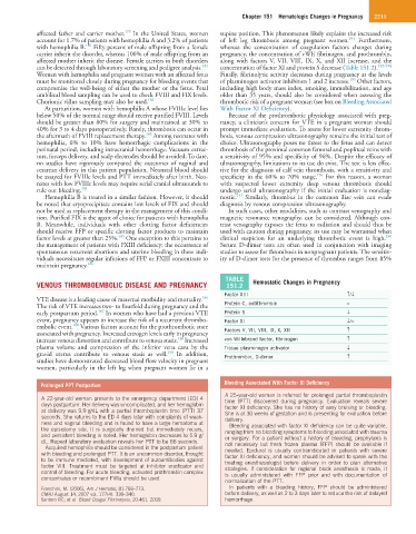
can be detected through laboratory screening and pedigree analysis. concentration of factor XI and protein S decrease (Table 151.2). 192–194
Women with hemophilia and pregnant women with an affected fetus Finally, fibrinolytic activity decreases during pregnancy as the levels
195
must be monitored closely during pregnancy for bleeding events that of plasminogen activator inhibitors 1 and 2 increase. Other factors,
compromise the well-being of either the mother or the fetus. Fetal including high body mass index, smoking, immobilization, and age
umbilical blood sampling can be used to check FVIII and FIX levels. older than 35 years, should also be considered when assessing the
Chorionic villus sampling may also be used. 182 thrombotic risk of a pregnant woman (see box on Bleeding Associated
At parturition, women with hemophilia A whose FVIIIc level lies With Factor XI Deficiency).
below 50% of the normal range should receive purified FVIII. Levels Because of the prothrombotic physiology associated with preg-
should be greater than 80% for surgery and maintained at 30% to nancy, a clinician’s concern for VTE in a pregnant woman should
40% for 3 to 4 days postoperatively. Rarely, thrombosis can occur in prompt immediate evaluation. To assess for lower extremity throm-
183
the aftermath of FVIII replacement therapy. Among neonates with bosis, venous compression ultrasonography remains the initial test of
hemophilia, 6% to 10% have hemorrhagic complications in the choice. Ultrasonography poses no threat to the fetus and can detect
perinatal period, including intracranial hemorrhage. Vacuum extrac- thrombosis of the proximal common femoral and popliteal veins with
tors, forceps delivery, and scalp electrodes should be avoided. To date, a sensitivity of 95% and specificity of 96%. Despite the efficacy of
no studies have rigorously compared the outcomes of vaginal and ultrasonography, limitations to its use do exist. The test is less effec-
cesarean delivery in this patient population. Neonatal blood should tive for the diagnosis of calf vein thrombosis, with a sensitivity and
196
be assayed for FVIIIc levels and PTT immediately after birth. Neo- specificity in the 60% to 70% range. For this reason, a woman
nates with low FVIIIc levels may require serial cranial ultrasounds to with suspected lower extremity deep venous thrombosis should
rule out bleeding. 184 undergo serial ultrasonography if the initial evaluation is nondiag-
197
Hemophilia B is treated in a similar fashion. However, it should nostic. Similarly, thrombus in the common iliac vein can evade
be noted that cryoprecipitate contains low levels of FIX and should diagnosis by venous compression ultrasonography.
not be used as replacement therapy in the management of this condi- In such cases, other modalities, such as contrast venography and
tion. Purified FIX is the agent of choice for patients with hemophilia magnetic resonance venography, can be considered. Although con-
B. Meanwhile, individuals with other clotting factor deficiencies trast venography exposes the fetus to radiation and should thus be
should receive FFP or specific clotting factor products to maintain used with caution during pregnancy, its use may be warranted when
196
185
factor levels at greater than 25%. One exception to this pertains to clinical suspicion for an underlying thrombotic event is high.
the management of patients with FXIII deficiency; the occurrence of Serum D-dimer tests are often used in conjunction with imaging
spontaneous recurrent abortions and uterine bleeding in these indi- studies to assess for thrombosis in nonpregnant patients. The sensitiv-
viduals necessitates regular infusions of FFP or FXIII concentrate to ity of D-dimer tests for the presence of thrombus ranges from 85%
maintain pregnancy. 186
TABLE
VENOUS THROMBOEMBOLIC DISEASE AND PREGNANCY 151.2 Hemostatic Changes in Pregnancy
Factor XIII ↑/↓
186
VTE disease is a leading cause of maternal morbidity and mortality.
The risk of VTE increases two- to fourfold during pregnancy and the Protein C, antithrombin =
187
early postpartum period. In women who have had a previous VTE Protein S ↓
event, pregnancy appears to increase the risk of a recurrent thrombo- Factor XI ↓/=
188
embolic event. Various factors account for the prothrombotic state Factors V, VII, VIII, IX, X, XII ↑
associated with pregnancy. Increased estrogen levels early in pregnancy
189
increase venous distention and contribute to venous stasis. Increased von Willebrand factor, fibrinogen ↑
plasma volume and compression of the inferior vena cava by the Tissue plasminogen activator ↓
190
gravid uterus contribute to venous stasis as well. In addition, Prothrombin, D-dimer ↑
studies have demonstrated decreased blood flow velocity in pregnant
women, particularly in the left leg when pregnant women lie in a
Prolonged PPT Postpartum Bleeding Associated With Factor XI Deficiency
A 25-year-old woman is referred for prolonged partial thromboplastin
A 22-year-old woman presents to the emergency department (ED) 4 time (PTT) discovered during pregnancy. Evaluation reveals severe
days postpartum. Her delivery was uncomplicated, and her hemoglobin factor XI deficiency. She has no history of easy bruising or bleeding.
at delivery was 9.9 g/dL with a partial thromboplastin time (PTT) 37 She is at 30 weeks of gestation and is presenting for evaluation before
seconds. She returns to the ED 4 days later with complaints of weak- delivery.
ness and vaginal bleeding and is found to have a large hematoma at Bleeding associated with factor XI deficiency can be quite variable,
the episiotomy site. It is surgically drained but immediately recurs, ranging from no bleeding symptoms to bleeding associated with trauma
and persistent bleeding is noted. Her hemoglobin decreases to 5.9 g/ or surgery. For a patient without a history of bleeding, prophylaxis is
dL. Repeat laboratory evaluation reveals her PTT to be 66 seconds. not necessary but fresh frozen plasma (FFP) should be available if
Acquired hemophilia should be considered in the postpartum patient needed. Epidural is usually contraindicated in patients with severe
with bleeding and prolonged PTT. It is an uncommon disorder, thought factor XI deficiency, and women should be advised to speak with the
to be immune mediated, with development of autoantibodies against treating anesthesiologist before delivery in order to plan alternative
factor VIII. Treatment must be targeted at inhibitor eradicator and strategies. If consideration for regional block anesthesia is made, it
control of bleeding. For acute bleeding, activated prothrombin complex is usually administered with FFP prior and with documentation of
concentrates or recombinant FVIIa should be used. normalization of the PTT.
Franchini, M. (2006), Am J Hematol, 81:768–773. In patients with a bleeding history, FFP should be administered
CMAJ August 14, 2007 vol. 177(4): 339–340. before delivery, as well as 2 to 3 days later to reduce the risk of delayed
Santoro RC, et al. Blood Coagul Fibrinolysis, 20:461, 2009. hemorrhage.