
Page 2579 - Hematology_ Basic Principles and Practice ( PDFDrive )
P. 2579
2302 Part XIII Consultative Hematology
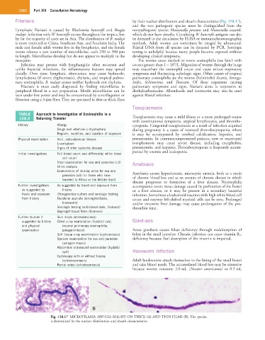
Filariasis by their nuclear distribution and sheath characteristics (Fig. 158.17),
and the two pathogenic species must be distinguished from the
Lymphatic filariasis is caused by Wuchereria bancrofti and Brugia nonpathogenic species Mansonella perstans and Mansonella ozzardi,
malayi. Infection with W. bancrofti occurs throughout the tropics, but which do not have sheaths. Circulating W. bancrofti antigens can also
by far the majority of cases are in Asia. The distribution of B. malayi be detected in the circulation by ELISA or immunochromatographic
is more restricted to China, Southeast Asia, and Southern India. The methods. Adult worms can sometimes be imaged by ultrasound.
male and female adult worms live in the lymphatics, and the female Filarial DNA from all species can be detected by PCR. Serologic
worm releases a vast number of microfilariae, each 250 to 300 µm testing is unhelpful because many people become exposed without
in length. Microfilariae develop but do not appear to multiply in the developing clinical symptoms.
mosquito. The worms cause marked or severe eosinophilia (see later) with
9
Infection may present with lymphangitis; often recurrent and counts greater than 1 × 10 /L. Migration of worms through the lungs
unlike bacterial infections, the inflammatory features may spread may exacerbate the eosinophil count and cause minor respiratory
distally. Over time, lymphatic obstruction may cause hydrocele, symptoms and fluctuating radiologic signs. Other causes of tropical
lymphedema (if severe elephantiasis), chyluria, and tropical pulmo- pulmonary eosinophilia are the worms (helminths) Ascaris, Strongy-
nary eosinophilia. B. malayi causes neither hydrocele nor chyluria. loides, Schistosoma, and Toxocara. Of these organisms causing
Filariasis is most easily diagnosed by finding microfilariae in pulmonary symptoms and signs, filariasis alone is responsive to
peripheral blood in a wet preparation. Motile microfilariae can be diethylcarbamazine. Albendazole and ivermectin may also be used
seen under low power and may be concentrated by centrifugation or against filarial infection.
filtration using a 3-µm filter. They are speciated in thin or thick films
Toxoplasmosis
TABLE Approach to Investigation of Eosinophilia in a
158.4 Returning Traveler Toxoplasmosis may cause a mild illness or a more prolonged course
with constitutional symptoms, atypical lymphocytes, and thrombo-
History Allergy cytopenia. Congenital toxoplasmosis as a result of infection acquired
Drugs and vitamins (L-tryptophan) during pregnancy is a cause of neonatal thrombocytopenia, where
Regions, localities, and duration of exposure it may be accompanied by cerebral calcification, hepatitis, and
Physical examination Skin, subcutaneous tissues pneumonitis. In immunocompromised patients, new or reactivated
Liver/spleen toxoplasmosis may cause severe disease, including encephalitis,
Signs of other systemic disease pneumonitis, and hepatitis. Thrombocytopenia is frequently accom-
panied by anemia and leukopenia.
Initial investigations Full blood count and differential white blood
cell count
Stool examination for ova and parasites (×3) Amebiasis
Urine analysis
Examination of midday urine for ova and Amebiasis causes hypochromic, microcytic anemia, both as a result
parasites (×3) (in those who have of chronic blood loss and as an anemia of chronic disease in which
traveled to Africa or the Middle East)
disease progresses to formation of a liver abscess. Neutrophilia
Further investigations As suggested by travel and exposure from accompanies severe tissue damage caused by perforation of the bowel
as suggested by history or a liver abscess, or it may be present in a secondary bacterial
travel and exposure Strongyloides culture and serologic testing infection. Sometimes a leukemoid reaction with high white blood cell
from history Duodenal aspirate (strongyloidiasis, count and extreme left-shifted myeloid cells can be seen. Prolonged
hookworm) and/or extensive liver damage may cause prolongation of the pro-
Serologic testing (schistosomiasis, filariasis) thrombin time.
Day/night blood films (filariasis)
Further studies if Skin snips (onchocerciasis)
suggested by history Chest x-ray examination (hydatid cyst, Giardiasis
and physical tropical pulmonary eosinophilia,
examination paragonimiasis) Acute giardiasis causes folate deficiency through malabsorption of
Soft tissue x-ray examination (cysticercosis) folate in the small intestine. Chronic infection can cause vitamin B 12
Sputum examination for ova and parasites deficiency because ileal absorption of the vitamin is impaired.
(paragonimiasis)
Abdominal ultrasound examination (hydatid
cyst) Hookworm Infection
Cystoscopy with or without biopsy
(schistosomiasis) Adult hookworms attach themselves to the lining of the small bowel
Rectal snips (schistosomiasis) and take blood meals. The accumulated blood loss may be extensive
because worms consume 2.0 mL (Necator americanus) or 0.5 mL
A B
Fig. 158.17 MICROFILARIA (BRUGIA MALAYI) ON THICK (A) AND THIN FILMS (B). The species
is determined by the nuclear distribution and sheath characteristics.