
Page 2607 - Hematology_ Basic Principles and Practice ( PDFDrive )
P. 2607
2320 Part XIII Consultative Hematology
CT scanning for identifying nodal disease and superior to both of Howell-Jolly bodies, which are nuclear remnants normally
lymphangiography and CT scanning at imaging the spleen. The removed by the spleen, is an excellent indicator of hyposplenism
availability of more sensitive, specific, and less toxic approaches to (Fig. 160.6). These are rarely seen until the spleen is largely non-
imaging permits more precise staging of Hodgkin lymphoma without functional or overwhelmed by other phagocytic functions, such as
the complications of splenectomy (Fig. 160.5). 12,13 extravascular hemolysis. Newborn infants commonly have visible
The role of PET scanning is expanding in other disorders as Howell-Jolly bodies, and splenic function appears to be at least
whole-body PET scanners and appropriate small-molecule markers somewhat impaired in the first week of life. Pappenheimer bodies
for individual diseases are more widely available. Imaging studies in (siderotic granules normally removed by the spleen) are often seen
small-animal models are very promising; splenic involvement can be in hyposplenic states, particularly when a component of hemolysis
identified with high sensitivity in mice. exists. Erythrocyte morphologic features reflect the lack of mem-
brane polishing by the spleen, with the presence of acanthocytes
and target cells. Granulocyte and platelet numbers are increased
TESTS OF SPLENIC FUNCTION during asplenic states, including splenic infarction and surgical
splenectomy.
The peripheral blood smear may be the most sensitive tool for To confirm suspected hyposplenism, the simplest test is a count of
identification of functional or anatomic hyposplenia. The presence pitted or pocked erythrocytes. Fixation in 0.5%–1.0% glutaraldehyde
A B
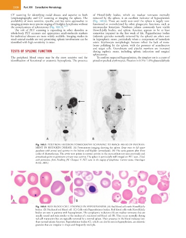
Fig. 160.5 POSITRON EMISSION TOMOGRAPHY SCANNING TO IMAGE AREAS OF INVOLVE-
MENT IN HODGKIN DISEASE. (A) Pretreatment imaging showing the spleen (large mass in left upper
quadrant with arrow) and activity in the kidney and bladder (arrowheads). (B) The same patient after three
cycles of chemotherapy. The arrow now points to normal activity in the myocardium not seen previously and
arrowheads point to persistent urinary tract activity. The spleen is particularly well imaged on PET scan. (Used,
with permission, from Friedberg JW, Chengazi V: PET scans in the staging of lymphoma: Current status, Oncologist
8:438, 2003.)
A B C
Fig. 160.6 RED BLOOD CELL FINDINGS IN HYPOSPLENISM. (A) Red blood cells with Howell-Jolly
bodies. (B) Nucleated red blood cell. (C) Cells with Pappenheimer bodies. Red blood cells with Howell-Jolly
bodies are seen in patients with hyposplenism. The cytoplasmic inclusions (A) are nuclear remnants that are
usually round and stain similar to the nucleus of a nucleated red blood cell (B). They occur normally during
red cell maturation but are typically removed by a normal spleen. Their presence in the blood indicates less-
than-normal splenic function. Pappenheimer bodies (C), which can also be seen in hyposplenism, are siderotic
granules that are irregular in shape and frequently multiple.