
Page 301 - Hematology_ Basic Principles and Practice ( PDFDrive )
P. 301
Chapter 23 Dendritic Cell Biology 249
+
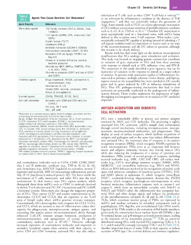
TABLE Agents That Cause Dendritic Cell Maturation a tolerization of T cells, such as when CD4 T-cell help is missing 38,39
23.1 or on activation by inflammatory cytokines in the absence of TLR
40
engagement, and they can potentially induce the generation of
Agent Property Molecules Tregs. Some stimuli, such as TSLP, can induce phenotypic maturation
Stimulatory agents TNF family members (TNF-α, CD40L, FasL, of DCs without concomitant secretion of proinflammatory cytokines
41
TRANCE) such as IL-12, IL-6, TNF-α, or IL-1. Therefore DC maturation is
TLR ligands (dsRNA, LPS, imiquimod, CpG more appropriately used in a functional sense, with mDCs being
ODNs) defined as able to prime naive T-cell responses. What makes a phe-
Growth factors (TSLP) notypically activated DC capable of priming instead of tolerizing a T
Interferons (IFN-α) cell appears multifactorial and dependent on factors such as the state
Adhesion molecules (CECAM-1 (CD66a) of the microenvironment and the DC subset in question, although
Costimulatory molecules (LIGHT, B7-DC) this remains to be clearly defined.
Receptors (FcR via Ag-Igs; TREM-2 via Recent work has shed some light on the intricate transcriptional
Dap-12) modifications that DCs undergo upon stimulation and activation.
Viruses or microbes (influenza, bacteria, This study was focused on mapping genetic variants that contribute
bacterial products) to variation of gene expression in DCs and how these associate
Chemokines (MCP, MIP1α, RANTES, IP10, with response to stimuli such as LPS, influenza virus, and IFN-β.
IL-8, MDC, TARC) The researchers in this study identified a gene signature that can be
Chemokine receptors (CCR7 and loss of CCR2 instructive of variation in the response to stimuli in a larger cohort
and CCR5) of samples. In genome-wide association studies of inflammatory dis-
eases such as psoriasis, multiple sclerosis, Crohn disease, and leprosy,
Inhibitory agents Drugs (rapamycin, FK506, cyclosporine A, regions closest to the susceptibility loci were enriched in DC-specific
dexamethasone, IVIg) genes and in genes induced by LPS or influenza stimulation of
Chemokines (IL-10) DCs. Thus DC pathogen-sensing mechanisms that lead to their
Viruses (EBV, vaccinia, canarypox, HSV) activation are potentially implicated in the pathogenesis of inflam-
Others (2 microglobulin) matory diseases. These studies underscore the importance of high-
Survival signals CD40L, TRANCE, B7-DC, Bcl-2 throughput and integrative approaches in the analysis of DC-mediated
Cell–cell interaction Activated cells (CD4 and CD8 cells [via responses. 42
CD40L])
NK cells, NK T cells ANTIGEN ACQUISITION AND DENDRITIC
+
Vδ1 , γδT cells
a Maturation is a complex process tightly linked to antigen acquisition and the CELL ACTIVATION
surrounding microenvironment. See text for more details.
Ag-Igs, Antigen–immunoglobulin immune complexes; Bcl-2, B-cell lymphoma DCs have a remarkable ability to process and present antigens
2; CCR, chemokine (C-C motif) receptor; CECAM-1, carcinoembryonic restricted by MHC and CD1 molecules. The processing is tightly
antigen-related cell adhesion molecule-1; CpG ODNs, CpG associated with DC activation. imDCs sample their environment
oligodeoxynucleotides; dsRNA, double-stranded RNA; EBV, Epstein-Barr virus;
FcR, Fc receptor; HSV, herpes simplex virus; IFN, interferon; IL, interleukin; through several mechanisms, including micropinocytosis, macropi-
IP10, interferon-γ–induced protein 10; IVIg, intravenous immunoglobulin; nocytosis, receptor-mediated endocytosis, and phagocytosis. They
LIGHT, homologous to lymphotoxins, exhibits inducible expression, and display an array of surface receptors, which facilitate acquisition of
competes with HSV glycoprotein D for herpesvirus entry mediator, a receptor antigens and pathogens and at the same time induce differentiation
expressed by T lymphocytes; LPS, lipopolysaccharide; MCP, macrophage/
monocyte chemotactic protein; MDC, macrophage and dendritic cell precursor; into activated DCs. An important class of receptors is the pattern
MIP1α, macrophage inflammatory protein 1α; NK, natural killer; RANTES, recognition receptors (PRRs), which recognize PAMPs expressed by
regulated on activation, normal T expressed and secreted; TARC, thymus and many microorganisms. PRRs serve as an important link between
activation-regulated chemokine; TNF, tumor necrosis factor; TRANCE, innate and adaptive immunity because they directly mature DCs
TNF-related activation-induced cytokine; TREM-2, triggering receptor expressed
on myeloid cells 2; TSLP, thymic stromal lymphopoietin. while also inducing the production of a variety of cytokines and
chemokines. PRRs consist of several groups of receptors, including
secreted molecules (e.g., MBL, CRP, SAP, LBP), cell surface mol-
and costimulatory molecules such as CD54, CD80, CD86, MHC ecules (e.g., CD14, macrophage mannose receptor [MMR], MSR,
43
class I and II molecules, cytokines (e.g., TNF-α, IL-12, IL-18), MARCO), and intracellular molecules (e.g., RIG-I and MDA5,
and chemokines (e.g., RANTES [regulated on activation, normal T which are RNA helicases involved in the recognition of nucleic acids
expressed and secreted], MIP-1α [macrophage inflammatory protein upon viral infection; stimulator of interferon genes [STING], DAI,
1α], IP-10 [interferon-γ–induced protein 10]). The latter enable the and AIM2 [absent in melanoma 2], which recognize intracellular
44
recruitment of T cells, monocytes, and other DCs into the local DNA ; nucleotide-binding oligomerization domain-like [NOD]
environment. In their mature state, DCs express markers, which receptors, which recognize peptidoglycan subcomponents or other
distinguish them from imDCs such as CD83 (a molecule involved bacterial molecules; inflammatory caspases, such as caspase-1 and
in thymic T-cell selection and DC–DC interactions) and DC-LAMP, caspase-5, which form an intracellular complex with NALP1 or
a lysosomal protein. Maturation also changes the migratory proper- NALP2 and NALP3 called the inflammasome that recognizes bac-
ties of DCs. They express CCR7 and acquire responsiveness to the terial RNA and other danger signals and induces the production
chemokines CCL19 and CCL21 that are expressed in the T-cell of the proinflammatory cytokines IL-1β and IL-18) (Table 23.2).
areas of lymph nodes where mDCs generate immune responses. TLRs, which constitute another group of PRRs, are expressed by
Concomitantly, DCs downregulate their receptors for CCL3, CCL4, imDCs and mediate activation by microbial components such as
and CCL5, which are secreted at sites of inflammation; reduce their peptidoglycan, LPS, flagellin, and unmethylated CpG DNA motifs.
capacity for phagocytosis, macropinocytosis, antigen uptake, and Ligation of the TLRs results in the activation of Rel family members,
processing; but acquire potent immunostimulatory ability through particularly the transcription factor nuclear factor κB (NFκB), c-Jun
enhanced T-cell–DC immune synapse formation, production of N-terminal kinase, and p38 mitogen-activated protein kinase, leading
immunoproteosomes, and upregulation of unique DC-specific to the initiation of the maturation process. 45,46 TLRs are unevenly
37
costimulatory molecules such as B7-DC. However, although distributed among DCs, with myeloid DCs expressing TLRs 2, 3, 4,
increased expression of costimulatory molecules and migration to 5, 7, and 8 and pDCs strongly expressing TLRs 7 and 9 (Table 23.3).
secondary lymphoid organs often correlate with their capacity to Another important feature of some TLRs is their capacity to induce
prime CD4 and CD8 immunity, activated DCs may also induce secretion of IFN type I for antiviral defense and immune regulation.