
Page 302 - Hematology_ Basic Principles and Practice ( PDFDrive )
P. 302
250 Part III Immunologic Basis of Hematology
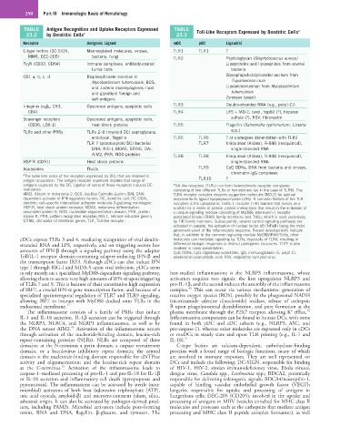
TABLE Antigen Recognition and Uptake Receptors Expressed TABLE Toll-Like Receptors Expressed by Dendritic Cells a
23.2 by Dendritic Cells a 23.3
Receptor Antigenic Ligand mDC pDC Ligand(s)
C-type lectins (DC-SIGN, Mannosylated molecules, viruses, TLR1 TLR1 ?
MMR, DEC-205) bacteria, fungi TLR2 Peptidoglycan (Staphylococcus aureus)
FcγR (CD32, CD64) Immune complexes, antibody-coated Lipoproteins and lipopeptides from several
tumor cells bacteria
CD1 a, b, c, d Bisphosphonate moieties in Glycophopshotidylinositol anchors from
Mycobacterium tuberculosis, BCG, Trypanosoma cruzi
and Listeria monocytogenes; lipid Lipoaminomannan from Mycobacterium
and glycolipid foreign and tuberculosis
self-antigens Zymosan (yeast)
Integrins (α v β 5 , CR3, Opsonized antigens, apoptotic cells TLR3 Double-stranded RNA (e.g., poly[I:C])
CR4) TLR4 LPS + MD-2, taxol, hsp60 (?), heparan
Scavenger receptors Opsonized antigens, apoptotic cells, sulfate (?), RSV, fibronectin
(CD36, LOX-1) heat shock proteins TLR5 Flagellin (Salmonella typhimurium, Listeria
TLRs and other PRRs TLRs 2–8 (myeloid DC) peptoglycans, spp.)
endotoxin, flagellin TLR6 TLR6 ? or undergoes dimerization with TLR2
TLR 7 (plasmacytoid DC) bacterial TLR7 Imiquimod (Aldara), R-848 (resiquimod),
DNA; RIG-I, MDA5, STING, DAI, single-stranded RNA
AIM2, PKR, NOD proteins TLR8 TLR8 Imiquimod (Aldara), R-848 (resiquimod),
HSP-R (CD91) Heat shock proteins single-stranded RNA
Aquaporins Fluids TLR9 CpG ODNs, DNA from bacteria and viruses,
chromatin-IgG complexes
a The table lists some of the receptors expressed by DCs that are involved in
antigen acquisition. The antigen receptor repertoire dictates that range of TLR10 ?
antigens captured by the DC. Ligation of some of these receptors induces DC a Toll-like receptors (TLRs) can form heterodimeric receptor complexes
maturation. consisting of two different TLRs or homodimers (as in the case of TLR4). The
AIM2, Absent in melanoma 2; BCG, bacillus Calmette-Guérin; DAI, DNA- TLR4 receptor complex requires supportive molecules (MD-2) for optimal
dependent activator of IFN-regulatory factors; DC, dendritic cell; DC-SIGN, response to its ligand lipopolysaccharide (LPS). A common feature of the TLR
dendritic cell–specific intercellular adhesion molecule-3-grabbing nonintegrin; receptors is the cytoplasmic Toll/IL-1 receptor (TIR) domain that serves as a
HSP-R, heat shock protein receptor; MDA5, melanoma differentiation- scaffold for a series of protein–protein interactions that result in the activation of
associated protein 5; NOD, nucleotide oligomerization domain; PKR, protein a unique signaling module consisting of MyD88; interleukin-1 receptor
kinase R; PRR, pattern recognition receptor; RIG-1, retinoid-inducible gene I; associated kinase (IRAK) family members; and Tollip, which is used exclusively
STING, stimulator of interferon genes; TLR, Toll-like receptor. by TIR family members. Subsequently, several central signaling pathways are
activated in parallel, the activation of nuclear factor κB (NFκB) being the most
prominent event of the inflammatory response. Recent developments indicate
that, in addition to the common signaling module MyD88/IRAK/Tollip, other
cDCs express TLRs 3 and 4, mediating recognition of viral double- molecules can modulate signaling by TLRs, especially of TLR4, resulting in
stranded RNA and LPS, respectively, and on triggering secrete low differential biologic responses to distinct pathogenic structures. TLR2 is also
amounts of IFN-β through a signaling pathway using the adaptor involved in cross-presentation.
CpG ODNs, CpG oligodeoxynucleotides; IgG, immunoglobulin G; poly(I:C),
Toll/IL-1 receptor domain-containing adapter-inducing IFN-β and polyinosinic-polycytidylic acid; RSV, respiratory syncytial virus.
the transcription factor IRF3. Although cDCs can also induce IFN
type I through RIG-I and MDA-5 upon viral infection, pDCs seem
to rely mostly on a specialized MyD88-dependent signaling pathway, best-studied inflammasome is the NLRP3 inflammasome, whose
allowing them to secrete very high amounts of IFN-α upon triggering activation requires two signals: the first upregulates NLRP3 and
of TLRs 7 and 9. This is because of their constitutive high expression pro-IL-1β, and the second induces the assembly of the inflammasome
48
of IRF7, a crucial IFN-α gene transcription factor, and because of a complex. This can occur via various mechanisms: generation of
specialized spatiotemporal regulation of TLR7 and TLR9 signaling, reactive oxygen species (ROS), possibly by the phagosomal NADH
allowing IRF7 to interact with MyD88 docked onto TLRs in the (nicotinamide adenine dinucleotide) oxidase, release of cathepsin
endosomal membrane. 47 B upon phagolysosomal destabilization, and pore formation at the
+
50
The inflammasome consists of a family of PRRs that induce plasma membrane through the P2X7 receptor, allowing K efflux.
IL-1 and IL-18 secretion. IL-1β secretion can be triggered through Inflammasome components can be found in human DCs, with some
the NLRP3, NLRC4, and NLRP1 inflammasomes, as well as by found in both pDC and cDC subsets (e.g., NLRP3, ASC, and
48
the DNA sensor AIM2. Activation of the inflammasome occurs pro-caspase-1), whereas other molecules are expressed only in cDCs
through activation of the nucleotide-binding domain, leucine-rich or moDCs in steady state and upon TLR priming (e.g., IL-1 and
repeat-containing proteins (NLRs). NLRs are composed of three IL-18). 51
domains: at the N-terminus a pyrin domain, a caspase recruitment C-type lectins are calcium-dependent, carbohydrate-binding
domain, or a baculovirus inhibitory repeat domain; the central proteins with a broad range of biologic functions, many of which
domain is the nucleotide-binding domain responsible for dNTPase are involved in immune responses. They are well represented on
activity and oligomerization; and the leucine-rich repeat domain DCs and include the following: DC-SIGN, responsible for binding
49
at the C-terminus. Activation of the inflammasome leads to of HIV-1, HIV-2, simian immunodeficiency virus, Ebola viruses,
caspase-1–mediated processing of pro-IL-1 and pro-IL-18 for IL-1β dengue virus, Candida spp., Leishmania spp.; BDCA2, potentially
or IL-18 secretion and inflammatory cell death (pyropoptosis and responsible for delivering tolerogenic signals; BDCA4/neuropilin-1,
pyronecrosis). The inflammasome can be activated by sterile (non- capable of binding vascular endothelial growth factor (VEGF);
microbial) activators of both host (adenosine triphosphate [ATP], langerin, responsible for uptake and processing of antigens in
uric acid crystals, amyloid-β) and microenvironment (alum, silica, Langerhans cells; DEC-205 (CD205), involved in the uptake and
asbestos) origin. It can also be activated by pathogen-derived prod- processing of antigens in MIIV (vesicles enriched for MHC class II
ucts, including PAMPs. Microbial activators include pore-forming molecules and proteases such as the cathepsins that mediate antigen
toxins, RNA and DNA, flagellin, β-glucans, and zymosan. The processing and MHC class II peptide complex formation), as well