Page 32 - Hematology_ Basic Principles and Practice ( PDFDrive )
P. 32

4     Part I  Molecular and Cellular Basis of Hematology


                       A                                                             B         C
                                                                3′ end
                                                                                3′    C:G   5′
                       5′ end                                 H   O                    A:T
                                        O                   H 2′   3′  H
                      5′ H C  O     N                             H                   G:C   5′    3′
                                                                                       A:T
                        2
                                                                     4′
                                                                                    C:G
                        4′  H  H  1′     N  H      N    N  1′  H  O  5′CH           T:A       T   A
                         H  3′  2′  H     O   H  N          N          2           G:C        A  T
                                                                                     T:A
                           O   H    CH 3                            O                 C:G     G  C
                                    Thymine      H      N                -             A:T
                        - O  P  O                      Adenine   O   P  O                     C  G
                                                                                       A:T
                           O           N      H            CH 3  H  O               C:G     3′    5′
                                                                                      G:C
                                              N  H    O       H  2′  3′ H           T:A
                        5′ H C  O   N                        1′  H  H   4′
                           2
                           4′  H  H  1′      N      H  N                           G T:A  C  5′
                                        N                   N     O   5′ CH 2  3′  G     C
                           H  3′  2′  H  Adenine         O  Thymine    O          C:G     G
                              O  H                                                A:U     T
                                                                                  T:A
                          - O  P  O                                 O  P  O -     C:G     A
                                                                                          G
                                          N              H          H  O          A:U     T
                              O                                                   A:U     T
                                                 O   H   N       H 2′   3′  H     T:A     A
                          5′ H C  O    N                        1′  H  H   4′     C:G     G
                             2
                             4′  H  H   1′     N  H       N                       G:C     C
                                          N                    N      O  5′ CH 2  G:C   A  C
                                                                                  T
                              H  3′  2′  H     N  H         O             O       A       A
                                                                                          T
                                 O  H   Guanine  H          Cytosine               A      T
                             - O  P  O                                 O  P  O -    T     A
                                                          H           H   O      5′    A  T  3′
                                 O                                  H  2′  3′ H
                                               O       H   N                           A:T
                             5′ H C  O      N                   N  1′  H  H   4′      G:C
                                2
                                                                                     C:G
                                4′  H  H  1′    N       H  N            O            T:A
                                                                    N      5′ CH 2
                                 H  3′  2′  H     N  H   O                          G:C
                                                                                     T:A
                                   O  H                         N          5′ end     C:G
                                - O  P  O  Cytosine           Guanine               5′  A:T  3′
                                  3′ end
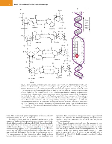
                        Fig. 1.1  STRUCTURE, BASE PAIRING, POLARITY, AND TEMPLATE PROPERTIES OF DNA. (A)
                        Structures of the four nitrogenous bases projecting from sugar phosphate backbones. The hydrogen bonds
                        between them form base pairs holding complementary strands of DNA together. Note that whereas A–T and
                        T–A base pairs have only two hydrogen bonds, C–G and G–C pairs have three. (B) The double helical structure
                        of DNA results from base pairing of strands to form a double-stranded molecule with the backbones on the
                        outside and the hydrogen-bonded bases stacked in the middle. Also shown schematically is the separation
                        (unwinding) of a region of the helix by mRNA polymerase, which is shown using one of the strands as a
                        template for the synthesis of an mRNA precursor molecule. Note that new bases added to the growing RNA
                        strand obey the rules of Watson–Crick base pairing (see text). Uracil (U) in RNA replaces T in DNA and,
                        like T, forms base pairs with A. (C) Diagram of the antiparallel nature of the strands, based on the stereochemi-
                        cal 3′  → 5′ polarity of the strands. The chemical differences between reading along the backbone in the
                        5′ → 3′ and 3′ → 5′ directions can be appreciated by reference to (A). A, Adenosine; C, cytosine; G, guanosine;
                        T, thymine; U, uracil.


        both). Most nucleic acid–synthesizing enzymes, for instance, add new   thymine at the same position of the opposite strand, or guanine with
        bases to the strand in a 5′ → 3′ direction.           cytosine—the Watson–Crick rules of base pairing. Two strands joined
           The ability of DNA molecules to store information resides in the   together in compliance with these rules are said to have “complemen-
        sequence of nucleotide bases arrayed along the polymer chain. Under   tary” base sequences.
        the physiologic conditions in living cells, DNA is thermodynamically   These  thermodynamic  rules  imply  that  the  sequence  of  bases
        most  stable  when  two  strands  coil  around  each  other  to  form  a   along one DNA strand immediately dictates the sequence of bases
        double-stranded  helix. The  strands  are  aligned  in  an  “antiparallel”   that must be present along the complementary strand in the double
        direction, having opposite 3′ → 5′ polarity (see Fig. 1.1). The DNA   helix. For example, whenever an A occurs along one strand, a T must
        strands are held together by hydrogen bonds between the bases on   be present at that exact position on the opposite strand; a G must
        one strand and the bases on the opposite (complementary) strand.   always be paired with a C, a T with an A, and a C with a G. In
        The  stereochemistry  of  these  interactions  allows  bonds  to  form   RNA–RNA  or  RNA–DNA  double-stranded  molecules,  U–A  base
        between the two strands only when adenine on one strand pairs with   pairs replace T–A pairs.
   27   28   29   30   31   32   33   34   35   36   37