
Page 35 - Hematology_ Basic Principles and Practice ( PDFDrive )
P. 35
Chapter 1 Anatomy and Physiology of the Gene 7
a
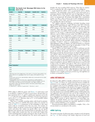
TABLE The Genetic Code Messenger RNA Codons for the decipher the exact encoding DNA sequence. These facts are summa-
1.1 Amino Acids rized by saying that the code is degenerate but not ambiguous.
Some specialized codons serve as punctuation points during
Alanine Arginine Asparagine Aspartic Acid Cysteine translation. The methionine codon (AUG), when surrounded by a
5′-GCU-3′ CGU AAU GAU UGU consensus sequence (the Kozak box) near the beginning (5′ end) of
the mRNA, serves as the initiator codon signaling the first amino
GCC CGC AAC GAC UGC acid to be incorporated. All proteins thus begin with a methionine
GCA CGA residue, but this is often removed later in the translational process.
Three codons, UAG, UAA, and UGA, serve as translation termina-
GCG AGA
tors, signaling the end of translation.
AGG The adaptor molecules mediating individual decoding events
Glutamic Acid Glutamine Glycine Histidine Isoleucine during mRNA translation are small (40 bases long) RNA molecules
called transfer RNAs (tRNAs). When bound into a ribosome, each
GAA CAA GGU CAU AUU tRNA exposes a three-base segment within its sequence called the
GAG CAG GGC CAC AUC anticodon. These three bases attempt to pair with the three-base
GGA AUA codon exposed on the mRNA. If the anticodon is complementary in
sequence to the codon, a stable interaction among the mRNA, the
GGG ribosome, and the tRNA molecule results. Each tRNA also contains
Leucine Lysine Methionine Phenylalanine Proline c a separate region that is adapted for covalent binding to an amino
acid. The enzymes that catalyze the binding of each amino acid are
UUA AAA AUG b UUU CCU constrained in such a way that each tRNA species can bind only to
UUG AAG UUC CCC a single amino acid. For example, tRNA molecules containing the
CUU CCA anticodon 3′-AAA-5′, which is complementary to a 5′-UUU-3′
(phenylalanine) codon in mRNA, can only be bound to or charged
CUC CCG
with phenylalanine; tRNA containing the anticodon 3′-UAG-5′ can
CUA only be charged with isoleucine, and so forth.
CUG Transfer RNAs and their amino acyl tRNA synthetases provide
for the coupling of nucleic acid information to protein information
Serine Threonine Tryptophan Tyrosine Valine
needed to convert the genetic code to an amino acid sequence.
UCU ACU UGG UAU GUU Ribosomes provide the structural matrix on which tRNA anticodons
UCC ACC UAC GUC and mRNA codons become properly exposed and aligned in an
orderly, linear, and sequential fashion. As each new codon is exposed,
UCA ACA GUA the appropriate charged tRNA species is bound. A peptide bond is
UCG ACG GUG then formed between the amino acid carried by this tRNA and the
AGU C-terminal residue on the existing nascent protein chain. The growing
chain is transferred to the new tRNA in the process, so that it is held
AGC
in place as the next tRNA is brought in. This cycle is repeated until
Chain Termination d completion of translation. The completed polypeptide chain is then
transferred to other organelles for further processing (e.g., to the
UAA
endoplasmic reticulum and the Golgi apparatus) or released into the
UAG cytosol for association of the newly completed chain with other
UGA subunits to form complex multimeric proteins (e.g., hemoglobin) and
a Note that most of the degeneracy in the code is in the third base position (e.g., so forth, as discussed in Chapter 5.
lysine, AA [G or C]; asparagine, AA [C or U]; valine, GUN [where N is any
base]).
b AUG is also used as the chain-initiation codon when surrounded by the Kozak mRNA METABOLISM
consensus sequence.
c Hydroxyproline, the 21st amino acid, is generated by posttranslational
modification of proline. It is almost exclusively confined to collagen subunits. In eukaryotic cells, mRNA is initially synthesized in the nucleus (see
d The codons that signal the end of translation, also called nonsense or Figs. 1.3 and 1.4). Before the initial transcript becomes suitable for
termination codons, are described by their nicknames amber (UAG), ochre translation in the cytoplasm, mRNA processing and transport occur
(UAA), and opal (UGA). by a complex series of events including excision of the portions of
A, Adenosine; C, cytosine; G, guanosine; T, thymine; U, uracil.
the mRNA corresponding to the introns of the gene (mRNA splic-
ing), modification of the 5′ and 3′ ends of the mRNA to render them
more stable and translatable, and transport to the cytoplasm. More-
DNA codon is called the genetic code (Table 1.1). Each amino acid over, the amount of any particular mRNA moiety in both prokaryotic
is encoded by a sequence of three successive bases. Because there are and eukaryotic cells is governed not only by the composite rate of
four code letters (A, C, G, and U), and because sequences read in the mRNA synthesis (transcription, processing, and transport) but also
5′ → 3′ direction have a different biologic meaning than sequences by its degradation by cytoplasmic ribonucleases (RNA degradation).
3
read in the 3′ → 5′ direction, there are 4 , or 64, possible codons Many mRNA species of special importance in hematology (e.g.,
consisting of three bases. mRNAs for growth factors and their receptors, proto-oncogene
There are 21 naturally occurring amino acids found in proteins. mRNAs, acute-phase reactants) are exquisitely regulated by control
Thus more codons are available than amino acids to be encoded. As of their stability (half-life) in the cytoplasm.
noted in Table 1.1, a consequence of this redundancy is that some Posttranscriptional mRNA metabolism is complex. Only a few
amino acids are encoded by more than one codon. For example, six relevant details are considered in this section.
distinct codons can specify incorporation of arginine into a growing
amino acid chain, four codons can specify valine, two can specify
glutamic acid, and only one each methionine or tryptophan. In no case mRNA Splicing
does a single codon encode more than one amino acid. Codons thus
predict unambiguously the amino acid sequence they encode. However, The initial transcript of eukaryotic genes contains several subregions
one cannot easily read backward from the amino acid sequence to (see Fig. 1.4). Most striking is the tandem alignment of exons and