
Page 36 - Hematology_ Basic Principles and Practice ( PDFDrive )
P. 36
8 Part I Molecular and Cellular Basis of Hematology
Intron
5 “CAP” GU AG GU AG GU AG (poly A tail)-3
5 UT 3 UT
Splice Splice
donor acceptor
site Splicing site
Protein coding sequence
5 “CAP” (poly A tail)-3
“CAP” site Poly (A) signal:
(1st base 5 - - - AUAA- - -AAAA(A)- - -3
transcribed)
Translation Termination Elements involved in
start site: AUG of translation: control of stability ~20 bp
UAG, UAA, (e.g., AU rich = unstable
or UGA codon mRNA)
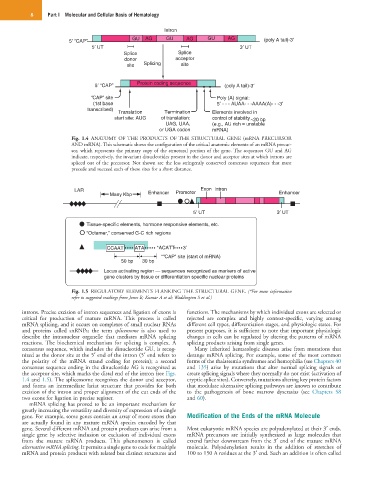
Fig. 1.4 ANATOMY OF THE PRODUCTS OF THE STRUCTURAL GENE (mRNA PRECURSOR
AND mRNA). This schematic shows the configuration of the critical anatomic elements of an mRNA precur-
sor, which represents the primary copy of the structural portion of the gene. The sequences GU and AG
indicate, respectively, the invariant dinucleotides present in the donor and acceptor sites at which introns are
spliced out of the precursor. Not shown are the less stringently conserved consensus sequences that must
precede and succeed each of these sites for a short distance.
LAR Exon Intron
Many Kbp Enhancer Promoter Enhancer
5 UT 3 UT
Tissue-specific elements, hormone responsive elements, etc.
“Octamer,” conserved G-C rich regions
CCAAT ATA *ACATT 3
*“CAP” site (start of mRNA)
50 bp 30 bp
Locus activating region — sequences recognized as markers of active
gene clusters by tissue or differentiation specific nuclear proteins
Fig. 1.5 REGULATORY ELEMENTS FLANKING THE STRUCTURAL GENE. (*For more information
refer to suggested readings from Jones B; Kumar A et al; Waddington S et al.)
introns. Precise excision of intron sequences and ligation of exons is functions. The mechanisms by which individual exons are selected or
critical for production of mature mRNA. This process is called rejected are complex and highly context-specific, varying among
mRNA splicing, and it occurs on complexes of small nuclear RNAs different cell types, differentiation stages, and physiologic states. For
and proteins called snRNPs; the term spliceosome is also used to present purposes, it is sufficient to note that important physiologic
describe the intranuclear organelle that mediates mRNA splicing changes in cells can be regulated by altering the patterns of mRNA
reactions. The biochemical mechanism for splicing is complex. A splicing products arising from single genes.
consensus sequence, which includes the dinucleotide GU, is recog- Many inherited hematologic diseases arise from mutations that
nized as the donor site at the 5′ end of the intron (5′ end refers to derange mRNA splicing. For example, some of the most common
the polarity of the mRNA strand coding for protein); a second forms of the thalassemia syndromes and hemophilia (see Chapters 40
consensus sequence ending in the dinucleotide AG is recognized as and 135) arise by mutations that alter normal splicing signals or
the acceptor site, which marks the distal end of the intron (see Figs. create splicing signals where they normally do not exist (activation of
1.4 and 1.5). The spliceosome recognizes the donor and acceptor, cryptic splice sites). Conversely, mutations altering key protein factors
and forms an intermediate lariat structure that provides for both that modulate alternative splicing pathways are known to contribute
excision of the intron and proper alignment of the cut ends of the to the pathogenesis of bone marrow dyscrasias (see Chapters 58
two exons for ligation in precise register. and 60).
mRNA splicing has proved to be an important mechanism for
greatly increasing the versatility and diversity of expression of a single
gene. For example, some genes contain an array of more exons than Modification of the Ends of the mRNA Molecule
are actually found in any mature mRNA species encoded by that
gene. Several different mRNA and protein products can arise from a Most eukaryotic mRNA species are polyadenylated at their 3′ ends.
single gene by selective inclusion or exclusion of individual exons mRNA precursors are initially synthesized as large molecules that
from the mature mRNA products. This phenomenon is called extend farther downstream from the 3′ end of the mature mRNA
alternative mRNA splicing. It permits a single gene to code for multiple molecule. Polyadenylation results in the addition of stretches of
mRNA and protein products with related but distinct structures and 100 to 150 A residues at the 3′ end. Such an addition is often called